
Image
|
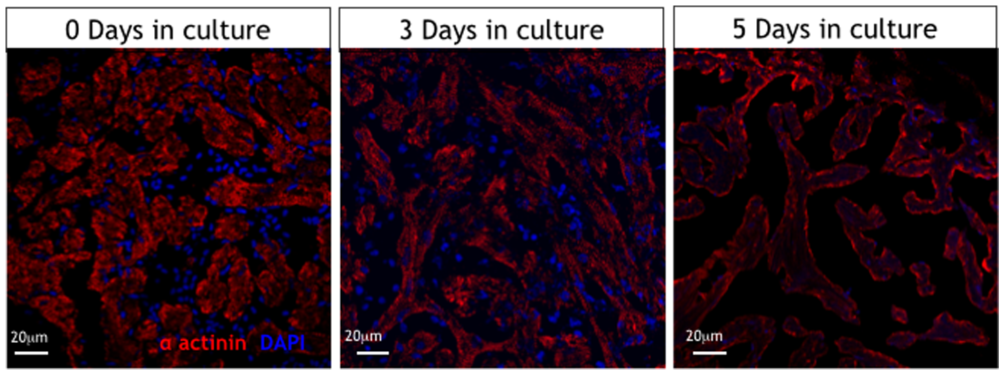
Figure Caption
Fig. 5
Immunohistology of isolated cultured hearts.
Alpha-actinin immunostaining showing well preserved sarcomere patterns in cardiomyocytes of cultured hearts at day 0 and day 3 with loss of this typical pattern by day 5 in culture. (red is sarcomeric alpha-actinin (mouse, clone EA-53, Abcam), blue is DAPI (Serva Heidelberg)).
Acknowledgments
This image is the copyrighted work of the attributed author or publisher, and
ZFIN has permission only to display this image to its users.
Additional permissions should be obtained from the applicable author or publisher of the image.
Full text @ PLoS One