
Image
|
Figure Caption
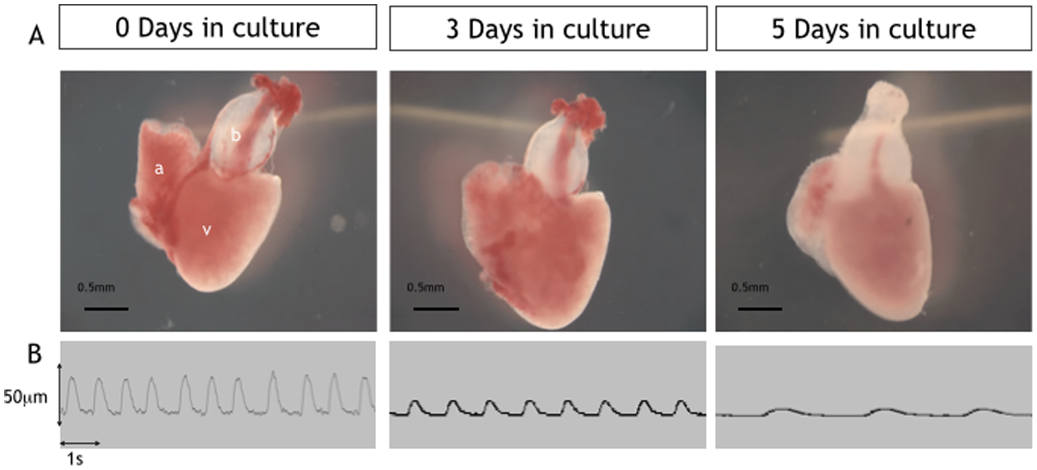
Fig. 1
Low power images of excised cultured hearts (days 0–5).
Upper panel (A) showing examples of excised hearts maintained in culture for 0, 3 and 5 days. Lower panel (B) shows example traces from Video-edge detection method displaying movement of the ventricle wall which was used to assess heart rate and ventricle function at days 0,3 and 5 in culture.
Acknowledgments
This image is the copyrighted work of the attributed author or publisher, and
ZFIN has permission only to display this image to its users.
Additional permissions should be obtained from the applicable author or publisher of the image.
Full text @ PLoS One