- Title
-
Post-transcriptional regulation of myotube elongation and myogenesis by Hoi Polloi
- Authors
- Johnson, A.N., Mokalled, M.H., Valera, J.M., Poss, K.D., and Olson, E.N.
- Source
- Full text @ Development
|
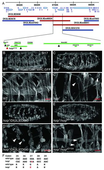
A mutation adversely affecting somatic muscle development maps to hoip. (A) The genomic region uncovered by Df(2L)ED690. Genes and direction of transcription are shown with blue arrows. Deficiencies that fail to complement hoip1 are shown in red; deficiencies that complement hoip1 are shown in dark blue. The minimal overlapping area among the deficiencies that fail to complement hoip1 contains eight genes. Of the four lethal transgene insertions (triangles) in the minimal overlapping area, only P{lacW}hoipk07104 (red triangle) failed to complement hoip1. (B-E) MHC.τGFP, Hand.nGFP expression in St17 embryos. (B) Wild-type embryos express membrane-localized τGFP in each somatic muscle in all embryonic segments. Somatic muscles are severely rounded (arrowheads) in hoip1 (C), hoip1/Df(2L)ED690 (D) and hoip1/P{lacW}hoipk07104 embryos (E). (B2-E2) High-magnification views of embryos shown in B-E. (F) hoip1 is a G37E missense mutation (see supplementary material Fig. S1I). In this and subsequent figures, embryos are oriented with anterior towards the left and dorsal towards the top. Coordinates refer to base pair positions on chromosome 2L. Scale bars: 20 μm. |
|
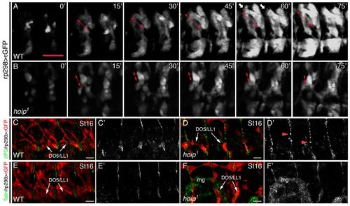
hoip embryos have myotube elongation defects. (A,B) Time-lapse images of rp298>τGFP embryos initiated at late St12. (A) Wild-type embryos showed robust myotube elongation at 30 minutes (double arrows) and developed extensive filopodia for attachment site recognition at 60 minutes (white arrows). (B) Myotubes established polarity in hoip1 embryos at 15 minutes but failed to elongate by 30 minutes. Polarized myofibers at 15 minutes compacted over time (double-headed arrows). (C,D) St16 rp298>τGFP embryos double labeled for GFP and βPS. (C) βPS localizes to myotendinous junctions in wild-type embryos. (D) Tendon cells express βPS in hoip1 embryos but localization is diffuse (red arrowheads). (C2,D2) βPS expression alone. (E,F) St16 rp298>τGFP embryos double labeled for GFP and Talin. Talin is expressed in tendon cells of wild-type (E) and hoip1 (F) embryos. (E2,F2) Talin expression alone. mg, midgut. Scale bars: 20 μm. |
|
Hoip regulates somatic muscle and cardioblast maturation but not precursor specification. (A,B) Mef2 and MHC protein expression in St16 embryos. Lateral views. Robust MHC and Mef2 expression is detectable in somatic muscles of hoip1/Cyo.lacZ embryos (A). Mef2 expression is unaffected in hoip1 embryos, whereas MHC is nearly absent from the somatic muscle (B). (C-F) St16 rp298.gal4>τ.GFP, rp298.nlacZ embryos double-labeled for GFP (red) and lacZ (green). (C-D2) Dorsal muscles. The number of lacZ+ nuclei is reduced in hoip1 embryos (C) compared with hoip1/Cyo.lacZ embryos (D); however, binucleated dorsal muscles show complete elongation (arrowheads). (E-F2) Lateral and ventral muscles. The number of lacZ+ nuclei is also reduced in lateral and ventral muscles in hoip1 embryos. Multinucleate lateral muscles show incomplete elongation (arrows). (G,H) Mef2 and MHC protein expression in St16 embryos. Dorsal views. (G) hoip1/Cyo.lacZ embryos express Mef2 and MHC in mature CBs. (H) hoip1 embryos express Mef2 but not MHC in a great majority of CBs. (I-L2) Mef2 and Tin protein expression. (I,J) hoip1/Cyo.lacZ embryos express Mef2 in all myogenic precursors, including CBs. Tin is expressed in four Mef2+ CBs per hemisegment at St13 (I; lateral view) and St16 (K; dorsal view). Mef2 and Tin expression in hoip1 CBs is comparable with control embryos at St13 (J) and St16 (L). (K2,L2) Tin expression alone. (M,N) High magnification micrographs of visceral muscles in St16 embryos. MHC expression is comparable between hoip1/Cyo.lacZ embryos (M) and hoip1 embryos (N). Both genotypes develop LVMs and CVMs in the visceral mesoderm. (O) hoip1 rp298>Hoip embryos express MHC protein at near wild-type levels in the somatic mesoderm. SM, somatic muscle; VM, visceral muscle; LVM, longitudinal visceral muscle; CVM, circular visceral muscle; CBs, cardioblasts. Open arrowheads in I,K show ectodermal cytoplasmic lacZ expression that distinguishes hoip1 heterozygotes from homozygotes. Scale bars: 20 μm. |
|
hoip is expressed in striated but not visceral muscle progenitors. (A) hoip gene organization and conservation within the Drosophila genus. The red line identifies genomic sequences used to generate the -225.nGFP and -225ΔE.nGFP hoip reporter genes. (B) St11 embryo labeled for hoip mRNA (green) and Mef2 (red). hoip mRNA is expressed in the Mef2-expressing cells of the somatic mesoderm, as well as in the fat body and the endoderm, but is absent from the neuroectoderm. (B2) hoip expression alone. (B2,B22) High magnification micrograph of the mesoderm shows hoip mRNA expression in the somatic but not the visceral mesoderm. (C-C2) At St13, hoip mRNA is still detectable in the developing somatic musculature. (D-E2) Hoip.-225.GFP embryos labeled for GFP (green) and Mef2 (red). GFP expression recapitulates hoip mRNA expression at St11 (D) and St13 (E). (F-G2) Hoip.-225ΔE.GFP embryos labeled for GFP (green) and Mef2 (red). GFP expression recapitulates hoip mRNA expression at St11 (F) but is undetectable at St13 (G). SM, somatic mesoderm; VM, visceral mesoderm; EN, endoderm. Scale bars: 20 μm. |
|
Hoip processes transcripts encoding sarcomere components. (A) Functional Gene Ontology (GO) analysis of misregulated transcripts in hoip1 embryos. Clusters of down- and upregulated transcripts are shown in red and green, respectively. The most significant cluster is associated with the term Contractile Fiber. (B) qPCR of Contractile Fiber transcript expression in hoip1 embryos compared with wild type. (C) The MHCemb transgene. The construct contains endogenous, somatic muscle MHC enhancer elements, multiple transcriptional start sites (colored 1st exons), an embryonic MHC cDNA and the endogenous poly A sites. bs, binding site. (D-F22) St16 embryos double-labeled for Tropomyosin (Tm) and MHC. Compared with hoip1/Cyo.lacZ embryos (D), both MHC and Tm are largely undetectable in the somatic and cardiac musculature of hoip1 embryos (E). In hoip1; MHCemb embryos, MHC protein expression is restored to near wild-type levels in somatic but not cardiac muscles; Tm remains largely undetectable in hoip1; MHCemb embryos (F). MHCemb does not rescue somatic muscle morphology defects (arrowheads) or MHC expression in cardioblasts (CBs). The Tm antibody recognizes both Tm1 and Tm2: RNA-seq showed a 0.50 (Tm1) and 0.09 (Tm2) fold change in hoip1 embryos compared with wild type (Table 2). (G,H) St16 embryos co-labeled for MHC mRNA and Hoechst. (G,G2) MHC mRNA shows both nuclear and cytoplasmic localization in the somatic muscle fibers of control embryos. (H,H2) MHC mRNA is exclusively detected in somatic muscle nuclei of hoip1 embryos. High magnification views in G2 and H2 show three segments of ventral oblique (VO) and ventral lateral (VL) muscles. (I) Quantification of MHC expression in the somatic musculature. Mean fluorescent intensity was calculated for lateral muscles over an entire segment (see supplementary material Fig. S7). The number of segments assayed is given for each genotype. Error bars represent s.e.m. Scale bars: 20 μm. |
|
Hoip is a conserved regulator of myogenesis. (A,B) St16 Drosophila embryos labeled for MHC protein. Compared with hoip1 embryos (A), hoip1 rp298>human NHP2L1 embryos show a significant restoration of MHC protein expression and muscle morphology in the somatic mesoderm (B). (C-E) Dorsal views of 14 hpf Tg (α-actin:GFP) zebrafish embryos. Embryos injected with control MO at the one-cell stage express robust GFP in somites (C, white arrowheads). Embryos injected with nhp2l1b ATG-MO (D) or 52UTR-MO (E) do not initiate GFP expression. ATG-MO and 52UTR-MO embryos develop a distinguishable neural tube by 14 hpf (black arrowheads). (F-G2) Dorsal view of 14 hpf Tg(α-actin:GFP) zebrafish embryos labeled for GFP and MF20 (which reacts with muscle MyHC isoforms). Embryos injected with control MO show robust GFP and MF20 expression in the somatic mesoderm (F), whereas ATG-MO injected embryos display little or no GFP or MF20 staining (G). (H) Dose-dependent response to nhp2l1b MOs. Percent penetrance was calculated as the number of embryos without detectable GFP fluorescence, relative to all injected embryos. Significance between ATG-MO/52UTR-MO and Cntrl MO was calculated using a t-test. **P<0.01, ***P<0.001. (I) qPCR of mesoderm transcript expression. Relative expression was calculated as mRNA levels in control versus ATG-MO-injected embryos after normalization to GAPDH. Error bars represent s.e.m. EXPRESSION / LABELING:
PHENOTYPE:
|
|
A forward genetic screen identified Hoip as a novel regulator of mesoderm development. (A) Crossing scheme to generate EMS mutants in a double GFP reporter background. Over 10,000 mutagenized genomes were screened. (B,C) MHC.τGFP, Hand.n-GFP expression in St17 embryos. Compared with wild-type embryos (B), P{lacW}hoipk07104 homozygous embryos (C) show severe muscle defects and apparent segmentation defects (white arrowheads). (D-F) St16 embryos stained with the PNS marker 22C10. Micrographs show four dorsal neuron clusters. The organization of the dorsal clusters (white arrows) and the pathway of the descending nerve (red arrows) are comparable among hoip1/Cyo (D), hoip1 ( |
|
Somatic muscles in hoip embryos extend filapodia in the direction of polarization. Time-lapse images of rp298. gal4>τGFP hoip1 somatic muscles beginning at late St12. Filapodia extend in the axis of myotube polarization (red arrowheads) and then retract. Scale bar: 10 μm. |
|
Muscle morphology and identity gene expression. (A, B) Nau expression in St12 embryos. The number of Nau+ nuclei is comparable between control and hoip1 embryos. (C, D) rp298.lacZ expression in St12 embryos. The number of lacZ+ nuclei is comparable between control and hoip1 embryos. (E, F) St16 rp298.gal4>τ.GFP, rp298.nlacZ embryos double-labeled for GFP (red) and lacZ (green). hoip1 embryos showed a significant reduction in the number of lacZ+ nuclei (F) compared with wild type (E). (G, H) Quantification of Nau+ and rp298.lacZ+ positive nuclei in the dorsal mesoderm. The number of segments quantified is given for each genotype and time point. Unpaired t-tests were performed to establish significance. Error bars represent s.e.m. (I, J) MHC.τGFP, Hand. nGFP expression in St16 embryos. (I) Wild-type and (J) MHC1 embryos show similar somatic muscle morphology. In particular LL1/DO5 muscles have elongated and attached in all segments (red arrows). ns, not significant; ***P<0.001. |
|
hoip expression during embryonic development. (A-E) Wild-type embryos hybridized with RNA probes antisense (A,C-E) or sense (B) to the hoip mRNA. (A,B) Post-cellularization blastoderm embryos. Fluorescent intensity is comparable between the antisense and sense probes. (C) St9 embryo co-labeled for hoip (green) and Mef2 (red). Weak hoip expression has initiated in the endoderm and mesoderm. (D,E) St14 (D) and St16 (E) embryos co-labeled for hoip (green) and MHC (red). hoip is expressed at high levels in the endoderm (en) and at lower levels in the somatic musculature (red arrowheads). (F) Genomic conservation 225 bp 52 to the hoip transcriptional start site. The highly conserved E-box sequence and mutations in Hoip.-225.DEGFP are shown. Genomic coordinates refer to base pair positions along chromosome 2L. |
|
Hoip localizes to both the nucleus and the cytoplasm. (A-D) Mef2>Hoip-HA embryos co-labeled for HA (green) and Tropomyosin (red). (A,B) Hoip is largely localized to the nucleus in St12 embryos, although some cytoplasmic staining is present. (C,D) Hoip is localized throughout the myofibers of St16 embryos. Enhanced localization is apparent in a subnuclear domain. |
|
Method to quantify MHC expression by mean fluorescence intensity. (A-C) Z-stacks including only the somatic muscles from St16 embryos stained for MHC. MHC expression was clearly restored in hoip1 embryos by MHCemb; however, muscle morphology was not (compare A with C). To quantify MHC expression in the somatic musculature (SM), we used the Zeiss LSM Zen 2011 software to trace the medial region of a segment using SBMs to define anterior-posterior position of each segment (white boxes). The software then calculated mean fluorescent intensity (MFI) within each boxed region. We also measured fluorescent intensity in the visceral musculature (VM). To normalize MHC expression, we divided SM MFI by VM MFI to obtain relative fluorescence for each segment. |
|
nhp2l1b is a Hoip orthologue and the nhp2l1b ATG-MO efficiently blocks translation. (A) Alignment between Hoip and zebrafish nhp2l1b protein sequences. Identical amino acids are shaded black; similar amino acids are shaded gray. (B) Alignment of the nhp2l1a/b translational start sequences with the ATG-MO and 5′UTR-MO. Target nucleotides are red. The beginning of the ORF is underlined. (C) Phylogenetic analysis of NHP2 and NHP2L1 proteins in worms (ce), flies (dm), zebrafish (dr) and humans (hs). Bootstrap values are given. (D) Zebrafish embryos co-injected with Control-MO and an eGFP construct containing the nhp2l1b target site in the 52UTR show robust eGFP expression at 12 hpf. (E) Embryos co-injected with ATG-MO and the same eGFP construct as in D show a significant reduction in eGFP expression. (F,G) Tg(α-actin:GFP) embryos injected with ATG-MO did not develop beyond 16 hpf. |


