
- Title
-
Zebrafish Ext2 is necessary for Fgf and Wnt signaling, but not for Hh signaling
- Authors
- Fischer, S., Filipek-Gorniok, B., and Ledin, J.
- Source
- Full text @ BMC Dev. Biol.

etv5b and pea3 expression is reduced in ext2 mutants. Lateral view (left panels) and dorsal view (right panels) of etv5b (A-D, M-P) and pea3 (E-L) expression in 38 hpf ext2 mutants (C, D, G, H, K, L, O, P) and WT embryos (A, B, E, F, I, J, M, N). (I-P) Embryos treated with 8 μM SU5402 3 h prior to fixation. Note that pea3 expression in WT embryos is only marginally affected by the inhibitor (compare E-F with I-J) while pea3 expression is almost completely inhibited by the same treatment in ext2 mutants (compare G-H with K-L). The arrows label the MHB. |
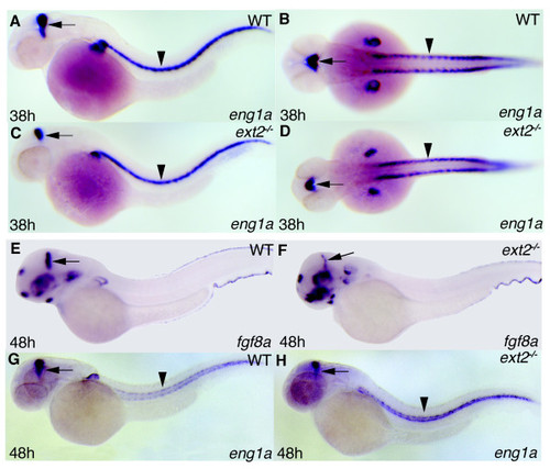
Reduced expression of eng1a and fgf8a in MHB but normal eng1a expression in muscle pioneers. Lateral view (A, C, E-H) and dorsal view (B, D) of 38 hpf (A-D) and 48 hpf (E-H) embryos. Expression of eng1a and fgf8a in ext2 mutants (C, D, F, H) and WT embryos (A, B, E, G). The arrows label the MHB and arrowheads label muscle pioneer cells. |
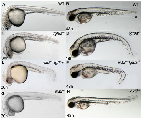
Tail development is disturbed in ext2;fgf8a double mutants. Lateral views of 30 hpf (left panels) and 48 hpf (right panels) embryos. (A, B) WT embryos, (C, D) fgf8a mutants and (E, F) fgf8a;ext2 double mutants. Tail morphology of ext2 mutants are indistinguishable from that of WT embryos at 30 hpf and 48 hpf (not shown). |
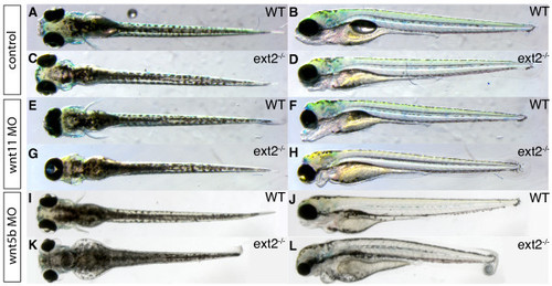
ext2 mutants are sensitive to a partial reduction in Wnt11 and Wnt5b translation. Dorsal view (left panels) and lateral view (right panels) of 3 dpf WT embryos (A-B, E-F, I-J) and ext2 mutants (C-D, G-H, K-L). Embryos injected with 10 ng MO1-wnt11 (E-H) and embryos injected with 7 ng MO1-wnt5b (I-L). Note that injection of the morpholinos results in a stronger phenotype in the ext2 mutants compared to WT embryos. |
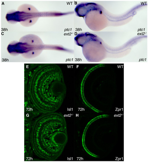
Hh signaling in ext2 mutants elicits normal ptc1 expression and functions normally in differentiation of cells in the retina. Lateral view (A, C) and dorsal view (B, D) of ptc1 expression in 38 hpf WT embryos (A, B) and ext2 mutants (C-D). Asterisks label the developing limbs. The difference in somite staining between A-B and C-D is within the range of individual variation (also see additional figure 3A-B) (E-H) Confocal sections of the retina at 72 hpf, with anterior to the top. Detection of the Isl1 protein (E, G) and Zpr1 protein (F, H) in WT retinas (E-F) and ext2 mutant retinas (G-H) reveal normal Hh signaling in ext2 mutants during patterning of the zebrafish retina. |
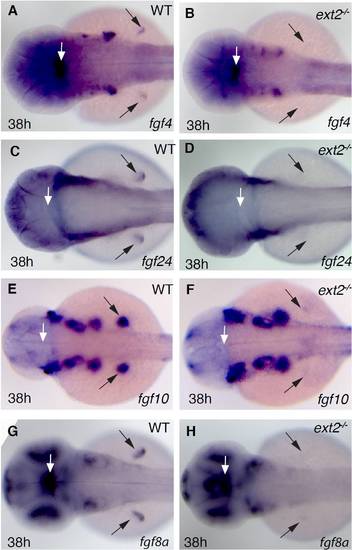
Expression of fgf genes in ext2 mutants. Dorsal view of fgf4 (A-B), fg24 (C-D), fgf10 (E-F), fgf8 (G-H) expression in control embryos (left panels) and ext2 mutants (right panels) at 38 hpf. Note that comparable levels of all examined fgf genes are expressed at 38 hpf, with the exception of the developing pectoral fin. Black arrows label the presence (left panels) or absence (right panels) of developing pectoral fins. |
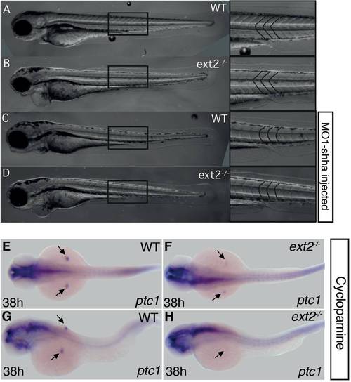
Hh signaling is not sensitizised in ext2 mutants. Morpholino injection experiment (A-D). Lateral view of 3 dpf control (A, C) and ext2 mutants (B, D). In (C, D) 14 ng MO1-shha have been injected in the one-cell stage which has resulted in U-shaped somites in a portion of injected embryos (see result section). The shape of the somites is emphasized in the right panel (A-D). Cyclopamine treatment experiment (E-H). ptc1 expression in 38 hpf embryos subjected to 50 μM cyclopamine 32-38 hpf (E-H). Dorsal view (E, F) and dorsolateral view (G-H). WT embryos (E, G) and ext2 mutants (F-H). |
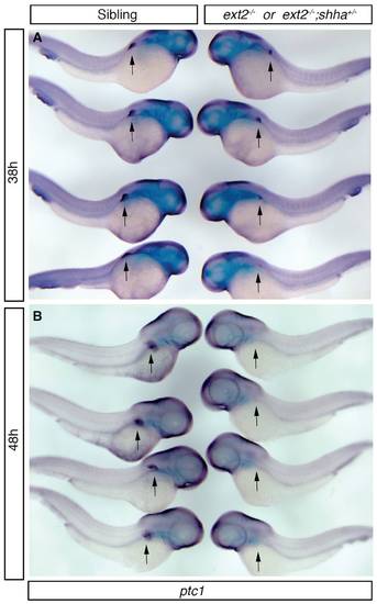
Hh signaling is not reduced in ext2-/-;shha+/- mutants. Genetic interaction experiment (A-B). Lateral view of ptc1 expression in 38 hpf (A) and 48 hpf (B) embryos from crossings of heterozygous ext2+/-;shha+/- double carriers with ext2+/- single carriers. Siblings (left row of embryos) and ext2-/-or ext2-/-;shha+/- mutants (right row of embryos). Arrows label the position of the pectoral fin. |