
|
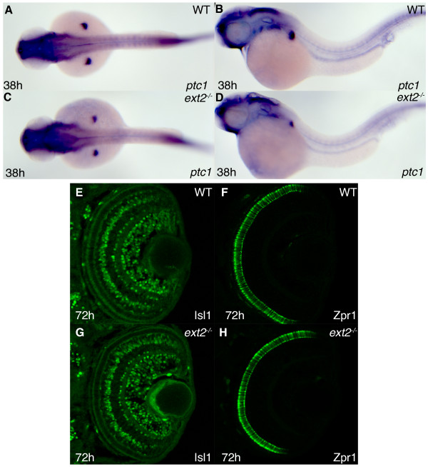
Fig. 5 Hh signaling in ext2 mutants elicits normal ptc1 expression and functions normally in differentiation of cells in the retina. Lateral view (A, C) and dorsal view (B, D) of ptc1 expression in 38 hpf WT embryos (A, B) and ext2 mutants (C-D). Asterisks label the developing limbs. The difference in somite staining between A-B and C-D is within the range of individual variation (also see additional figure 3A-B) (E-H) Confocal sections of the retina at 72 hpf, with anterior to the top. Detection of the Isl1 protein (E, G) and Zpr1 protein (F, H) in WT retinas (E-F) and ext2 mutant retinas (G-H) reveal normal Hh signaling in ext2 mutants during patterning of the zebrafish retina.