- Title
-
Delaying gal4-driven gene expression in the zebrafish with morpholinos and gal80
- Authors
- Faucherre, A., and López-Schier, H.
- Source
- Full text @ PLoS One
|
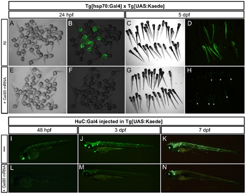
Gal80 expression inhibits Gal4 activity. (A–H) Embryos resulting from a cross between Tg[hsp70:Gal4] and Tg[UAS:Kaede] fish were either non-injected (NI, A–D) or injected with 100 pg of mRNA encoding full-length Gal80 (E–H). Representative specimens are depicted at 24 hpf (A–B, N = 18 GFP+/72 fish; E–F, N = 0 GFP+/75 fish) and 5 dpf (C–D, N = 18 GFP+/77 fish; G–H, N = 19 GFP+/82 fish). In H, asterisks indicate the fish displaying GFP. (I–N) Embryos resulting from a cross of Tg[UAS:Kaede] fish were injected with a DNA encoding full-length Gal4 under the control of the HuC promoter either alone (NI, I–K, N = 32) or with 100 pg of mRNA encoding full-length Gal80 (L–N, N = 37). Representative specimens are depicted at 48 hpf, 3 and 7 dpf. |
|
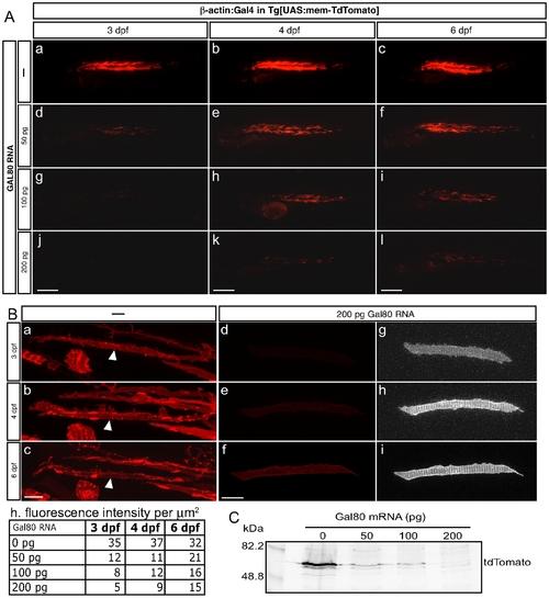
Gal80 RNA temporarily inhibits Gal4 activity in a dose-dependant manner. (A) Embryos resulting from a cross of Tg[UAS:mem-TdTomato] fish were injected with a DNA encoding full-length Gal4 under the control of the β-actin promoter alone (a–c) or with 50 pg (d–f, N = 21), 100 pg (g–i, N = 23) or 200 pg (j–l, N = 20) of mRNA encoding full-length Gal80. Representative specimens are depicted at 3, 4 and 6 dpf. (Scale bars: 300 μm). (B) Red fluorescence quantification of tdTomato expressing cells. (a–c) Maximal projection of muscle fibers from a embryo resulting from a cross of Tg[UAS:mem-TdTomato] fish injected with the β-actin:Gal4 construct alone. The white arrowheads indicate the quantified cell. (d–f) Maximal projection of muscle fibers from a embryo resulting from a cross of Tg[UAS:mem-TdTomato] fish injected with the β-actin:Gal4 construct and 200 pg of mRNA encoding full-length Gal80. Z-stacks have been captured at the same settings as in a–c. (g–i) Same as d-f with over-exposure. (Scale bars: 20 μm) (h) Quantitative values of fluorescence per μm2. Each value corresponds to the average of the mean values obtained for three different regions of each cell. (C) Anti-tdTomato western blotting of 3dpf fish resulting from a cross of Tg[UAS:mem-TdTomato] fish injected with the β-actin:Gal4 construct alone or with 50, 100 or 200 pg of mRNA encoding full-length Gal80. |
|
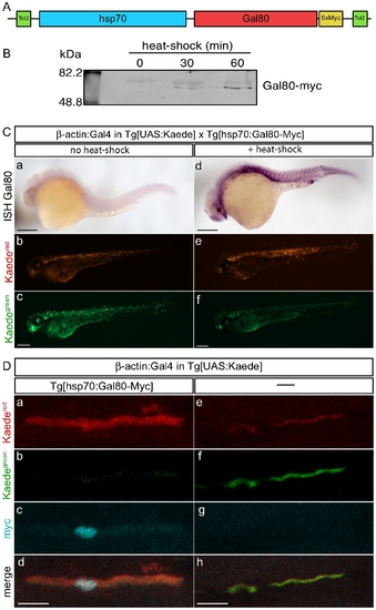
Temporal control of Gal80 expression. (A) Schematic representation of the construct hsp70:Gal80-myc. (B) Anti-myc western blotting of 48 hpf fish resulting from a cross of Tg[hsp70:Gal80-myc] fish after 0, 30 and 60 minutes of heat-shock. (C) Embryos resulting from a cross between Tg[hsp70:Gal80-Myc] and Tg[UAS:Kaede] fish were not submitted (a–c) or submitted to a one-hour heat-shock (d–f). Representative specimens are depicted for anti-Gal80 in situ hybridization at 30 hpf (a, d) or for Kaede expression at 2 dpf (b–c, d–e). (Scale bars: 150 µm). (D) Embryos resulting from a cross between Tg[hsp70:Gal80-Myc] and Tg[UAS:Kaede] fish were injected with a DNA encoding full-length Gal4 under the control of the β-actin promoter. At 24 hpf, the embryos were subjected to a 30-minute heat-shock at 39 degrees and immediately photo-converted. The following day, immunostaining anti-myc (c, g) was performed on fish expressing Gal80 (a–d) or not expressing Gal80 (e–h). The figure depicts muscle fibers for the expression of Kaedered (a, e) and Kaedegreen (b, f). (Scale bars: 20 μm). |
|
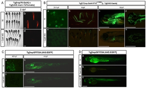
Gal4 morpholino temporarily inhibits Gal4 expression in Gal4-VP16 lines. (A) Embryos resulting from a cross between Tg[hsp70:Gal4] and Tg[UAS:mem-TdTomato] fish were either non-injected (NI, Aa-b, N = 27) or injected with 3 ng of Gal4 MO (Ac-d, N = 37). Representative specimens are depicted at 2 dpf. (B) Embryos resulting from a cross of Tg[ET(hsp:Gal4VP16s1006t);UAS:Kaede] double transgenic animals were either non-injected (NI, Ba-d, N = 47) or injected with 3 ng of Gal4 MO (Be-h, N = 52). Representative specimens are depicted at 24 hpf (Ba, e), 4 dpf (Bb, f) and 6 dpf (Bc-d, Bg-h). (C) Embryos resulting from a cross of Tg[hspGFF53A;UAS:EGFP] double transgenic animals were either non-injected (NI, Ca-b, N = 64) or injected with 3 ng of Gal4 MO (Cc-d, N = 58). Representative specimens are depicted at 48 hpf (Ca, c) and 4 dpf (Cb, d). (D). Embryos resulting from a cross of Tg[hspGFF53A;UAS:EGFP] double transgenic animals were either non-injected (NI, Da), injected with 5 ng of Gal4 MO (Db) or injected with 5 ng of a control MO (Dc). Representative specimens are depicted at 48 hpf. (Scale bars: 600 μm for A; 300 μm for B and C, 150 μm for D). |
|
Gal4 morpholino temporarily inhibits Gal4 expression in a dose-dependant manner. Embryos resulting from a cross of Tg[hspGFF53A;UAS:EGFP] double transgenic animals were either non-injected (A–E) or injected with 1 ng (F–J), 3 ng (K–O) or 5 ng (P–T) of Gal4 MO (N = 94). Representative specimens are depicted at 2, 4 and 6 dpf. White arrows indicate green fluorescence at the level of the posterior afferent lateralis ganglion. (Scale bars: 150 μm). |
|
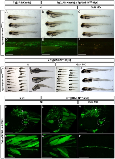
Gal4 morpholino allows bypassing of early developmental stages. (A–F) Embryos resulting from an incross between Tg[ET(hsp:Gal4VP16s1001t);UAS:Kaede] double trangenics (A,D) or from Tg[ET(hsp:Gal4VP16s1001t);UAS:Kaede] with Tg[UAS:NICD-Myc] animals (B–C, E–F) were either non-injected (NI, A–B, D–E) or injected with 3 ng of Gal4 MO (C,F). Representative specimens are depicted at 3 dpf. The lower panel shows a magnification of the different specimens after DiASP treatment (D–F). (G–J) Embryos resulting from a cross between Tg[ET(hsp:Gal4VP16s1006t)] and Tg[UAS:NICD-Myc] animals were either non-injected (G and magnification in H) or injected with 3 ng of Gal4 MO (I and magnification in J). Representative specimens are depicted at 6 dpf. (K–P) Embryos resulting from a cross between Tg[hspGFF53A;UAS:EGFP] double transgenic animals and either wt (K, N) or Tg[UAS:NICD-Myc] (L–M, O–P) animals were either non-injected (K–L, N–O) or injected with 3 ng of Gal4 MO (M, P). Representative specimens are depicted at 7 dpf. (K–M) Maximal projections of posterior lateralis afferent ganglion (dashed circles) and central projection (white arrowheads). (N–P) Maximal projections of posterior lateralis afferent nerve. (Scale bars: 150 μm for A–F, H and J; 300 μm for G and I; 100 μm for K–P). EXPRESSION / LABELING:
PHENOTYPE:
|

