
- Title
-
The predominant protein arginine methyltransferase PRMT1 is critical for zebrafish convergence and extension during gastrulation
- Authors
- Tsai, Y.J., Pan, H., Hung, C.M., Hou, P.T., Li, Y.C., Lee, Y.J., Shen, Y.T., Wu, T.T., and Li, C.
- Source
- Full text @ FEBS J.

Spatial and temporal expression of prmt1 by WISH and immunofluorescent analysis. Zebrafish embryos at the one-cell stage (A), two-cell stage (B), four-cell stage (C), 6 hpf (D), 12 hpf (E, F), 24 hpf (G), 48 hpf (H) and 72 hpf (I) were analyzed by WISH. A dorsal view of the 12 hpf is shown in (F). WISH with sense riboprobe is shown in (J). Immunofluorescent analysis with anti-PRMT1 of 24-hpf embryos is shown in (K). a, adaxial cells; e, eye; f, forebrain; h, hindbrain; m, midbrain; mhb, mid-hindbrain boundary; ov, otic vesicle; som, somites. |
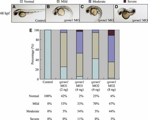
Defective phenotypes in prmt1 knockdown zebrafish. Phenotypes of embryos injected with zprmt1 MO at 48 hpf. Uninjected wild-type embryos are shown (A). The injected embryos are classified into mild, moderate and severe according to the phenotypes at 48 hpf. The three types of MO injected embryos at 48 hpf are shown in (B–D). The injected embryos with normal body axes as the wild-type are classified as ‘normal’. (E) Frequencies of three phenotypes caused by injection of prmt1 MO (2 or 4 ng of MO1 and 4, or 8 ng of MO2). The injected embryos with normal body axes as the wild-type are classified as ‘normal’. PHENOTYPE:
|

Reduced PRMT1 protein expression, type I protein arginine methyltransferase activity and specific protein arginine methylation in prmt1 morphants. (A) Proteins were prepared from embryos either not injected, or injected with prmt1 MO1. Western blot analysis of PRMT1 protein in embryos injected with 4 ng of MO1 with phenotypes classified as mild or moderate at 48 hpf are shown. Detection by anti-β-actin was used as a loading control. WT, wild-type; M, morphants. (B) In vitro methylation was conducted with extracts from MO2 (8 ng) injected embryos at 24, 48 and 72 hpf as the source of protein arginine methyltransferase and recombinant mouse fibrillarin as the methyl-accepting protein. The samples were separated by SDS/PAGE and the methylated proteins were detected by fluorography. (C) Arginine-methylated proteins in 48 hpf embryos were detected by western blotting with an asymmetric dimethylarginine-specific antibody ASYM24. Detection by anti-β-actin was used as a loading control. (D) Western blot analysis of H4R3me2 levels in 48 hpf morphant embryos. Analysis of histone H4 served to normalize levels of H4R3me2 in morphants and wild-type embryos. PHENOTYPE:
|
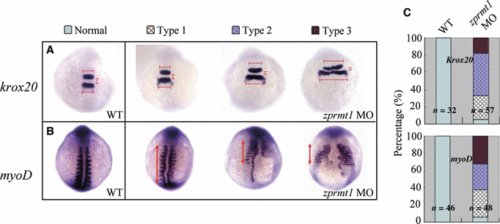
Defective phenotypes at segmentation stage for prmt1 morphants. (A) Dorsal view of embryos (10-somite stage) for krox20 staining. The positions of r3 and r5 are indicated. The widths of r3 and r5 and the vertical distance between r3 and r5 are indicated. In type 1 morphants, r3 was almost normal but r5 was slightly extended laterally. The width of r3 and r5 were extended to < 1.5-fold in type 2 and even to 2–2.5-fold in type 3. (B) Expression of myoD at paraxial/adaxial mesoderm at the 10-somite stage. Dorsal views, anterior at top. The lengths of myoD expressed paraxial/adaxial mesoderm are indicated. In type 1, 2 and 3 morphants, the length was < 0.8–0.9, 0.6–0.7 and 0.5 compared to normal. (C) The phenotypes were classified according the degree of abnormality as type 1, 2, and 3. Percentages of wild-type and morphants embryos within each phenotypic category are shown in the bar graphs. n, total embryos counted in the experiments. EXPRESSION / LABELING:
PHENOTYPE:
|

Knockdown of prmt1 induces gastrulation defects. Wild-type and prmt1 MO2 (8 ng) injected embryos at 10 hpf were examined. (A) The morphants showed abnormal morphology at the end of epiboly (10 hpf) as reflected by different degrees of open blastopores. Dashed arrows and semicircles depict embryo lengths and angles between anterior–posterior ends. Lateral views, dorsal to the right. (B) ntl (staining the forerunner cell group, axial chorda mesoderm) staining of the embryos at 10 hpf. Dorsal views, anterior at top. The morphants are grouped according to the degree of shortening and widening of the notochord. PHENOTYPE:
|
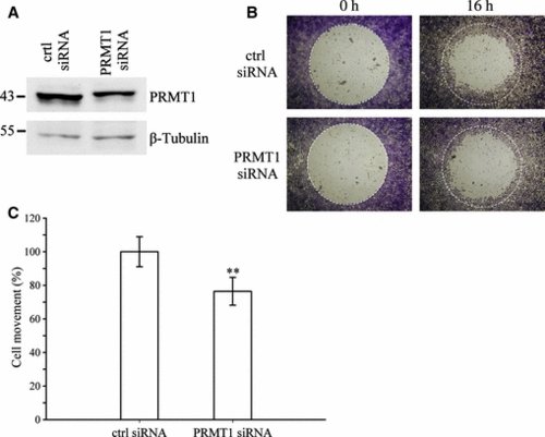
Reduced cell migration in a prmt1-deficient cell model. Huh7 cells were treated with control or prmt1 siRNA. (A) Cell extracts from the siRNA-treated cells were immunoblotted with anti-PRMT1. Detection by anti-β-tubulin was used as a loading control. Reduced PRMT1 protein expression by siRNA was normalized with the β-tubulin signal. (B) Images of the pre-migration and post-migration cells stained with crystal violet are shown. The white circles indicate areas covered by the stoppers before cell migration. (C) Quantification of cell movement is represented as the percentage of the area covered by migrated cells in prmt1 siRNA-treated cells compared to that in control cells. Data are shown as the mean ± SD of two independent experiments performed in quadruplicate. A statistically significant difference between the two siRNA-treated cells is indicated (**P < 0.01; Student’s t-test). |
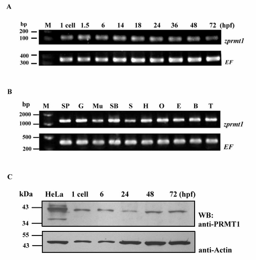
Expression of zebrafish prmt1 mRNA and protein during early development. (A) The expression of prmt1 during different zebrafish developmental stages analyzed by RT-PCR. (B) The expression of prmt1 in different zebrafish adult tissues analyzed by T-PCR. EF indicated the RT-PCR product of elongation factor 1A as a loading control. hpf, hours post fertilization; SP: spleen, S: skin, E: eye, G: gill, H: heart, B: brain, Mu: muscle, O: ovary, T: testis, SB: swim bladder. (C) Endogenous PRMT1 protein (approximately 42 kDa) in extracts of different embryonic stages (1 cell, 6, 24, 48 and 72 hpf) was detected by Western blot analysis. Lane 1: HeLa cell extract was included as a positive control. Detection by anti-β-actin was used as a loading control. |

Defective phenotypes in prmt1 knock-down zebrafish at 72, 96 or 120 hpf. Phenotypes of embryos injected with prmt1 MO at 72, 96 or 120 hpf. Un-injected wild-type embryos are shown at 72 (A), 96 (E) and 120 hpf (I). The injected embryos are classified into mild, moderate and severe. The three types of MO injected embryos at 72 hpf are shown in (B, C, D), at 96 hpf are shown in (F, G, H), at 120 hpf are shown in (J, K, L). PHENOTYPE:
|
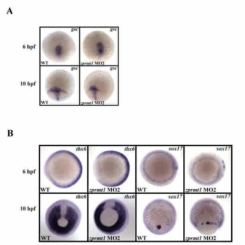
Knockdown of prmt1 induces gastrulation defects. Wild-type and prmt1 MO2 (8 ng) injected embryos at 6 or 10 hpf (the end of epiboly) were examined. (A) gsc (mesendodermal marker) staining of embryos of 6 or 10 hpf. Dorsal view. (B) tbx6 and sox17 staining of embryos at 6 or 10 hpf. For both markers at 6 hpf, top view dorsal to the right. For tbx6 staining at 10 hpf, bottom view dorsal to the right. For sox17 at staining at 10 hpf, lateral view. |