Fig. 4
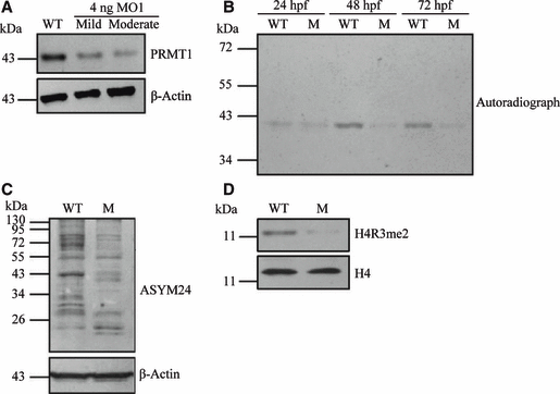
Reduced PRMT1 protein expression, type I protein arginine methyltransferase activity and specific protein arginine methylation in prmt1 morphants. (A) Proteins were prepared from embryos either not injected, or injected with prmt1 MO1. Western blot analysis of PRMT1 protein in embryos injected with 4 ng of MO1 with phenotypes classified as mild or moderate at 48 hpf are shown. Detection by anti-β-actin was used as a loading control. WT, wild-type; M, morphants. (B) In vitro methylation was conducted with extracts from MO2 (8 ng) injected embryos at 24, 48 and 72 hpf as the source of protein arginine methyltransferase and recombinant mouse fibrillarin as the methyl-accepting protein. The samples were separated by SDS/PAGE and the methylated proteins were detected by fluorography. (C) Arginine-methylated proteins in 48 hpf embryos were detected by western blotting with an asymmetric dimethylarginine-specific antibody ASYM24. Detection by anti-β-actin was used as a loading control. (D) Western blot analysis of H4R3me2 levels in 48 hpf morphant embryos. Analysis of histone H4 served to normalize levels of H4R3me2 in morphants and wild-type embryos.

