
- Title
-
Zebrafish notch signalling pathway mutants exhibit trunk vessel patterning anomalies that are secondary to somite misregulation
- Authors
- Therapontos, C., and Vargesson, N.
- Source
- Full text @ Dev. Dyn.

Expression of deltaC, deltaD, and notch1a at 24hpf. A-C: Expression of deltaC (A), deltaD (B), and notch1a (C) in 24hpf embryos. Note strong expression in head and somites. Scale bar = 200 μm. |

ISV defects in aei, bea, and des Notch mutant embryos at 24, 48, 72, and 96 hpf. A–P: Fluorescence images of the middle portion of the zebrafish embryo as indicated in Q. TG(fli1:egfp)y1 (A,E,I,M); aei (B,F,J,N), bea (C,G,K,O), and des (D,H,L,P) embryos at 24 (A-D), 48 (E–H), 72 (I-L), and 96 (M-P) hpf. The vascular patterning defects do not recover over time. The regularity of ISV sprouting in control embryos is highlighted by white arrowheads (A,E,I,M). Mutant ISVs show irregular spacing (white arrowheads), abnormal paths (white arrows), and aberrant branching patterns (white asterisks). The positions of the DA, PCV, ISVs, and DLAV are indicated. Scale bar = 100 μm. |

Anterior somites in the Notch mutants are normal. Bright field (A-D) and myoD expression (E-H) in wildtype (A, E) or Notch mutant (B-D, F–H) at 24hpf. Somite boundaries are normal in anterior but not posterior somites. Black arrowheads highlight normal somites. In aei, the first 8 somites form normally; in bea, the first 3 form normally; and in des, the first 7 form normally. Scale bar = 200 μm. |
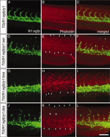
ISVs follow the irregular somite boundaries in Notch mutants. Wildtype (A-C) and Notch mutant embryos, aei (D–F), bea (G-I), and des (J-L), were stained with phalloidin to reveal somitic actin fibres (in red; B,E,H,K). The ISV vascular pattern revealed by the green fluorescence of the fli1:EGFP embryos (A,D,G,J) is seen correlating with the somitic boundaries (B,E,H,K) in the merged images (C,F,I,L). Asterisks indicate misplaced ISV over somite. Scale bar = 100 μm. |
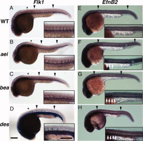
Expression of vascular markers in Notch mutant embryos is undisturbed at 24 hpf. Expression of Flk1 (A-D) in the zebrafish trunk reveals expression in vessels in control (A) and the aei, bea, and des Notch mutant embryos (B-D), even in the disrupted posterior somites (between black arrowheads). The anterior somites are normal (black asterisk). The disorganised ISVs are shown at higher magnification, inset. Expression of Efnb2 an arterial marker (E) is also present in each of the mutants (F-H), but in an irregular pattern in posterior somites (between black arrowheads; shown at higher magnification, inset). Efnb2 is expressed in a normal pattern in anterior somites of each mutant (white arrows, inset; F-H). Scale bar = 200 μm. |

Expression of Sema3a2 and plexinD1 in Notch mutant embryos. A-H: 24 hpf wildtype (A,E) and Notch mutant embryos, aei (B,F), bea (C,G), des (D,H) were analysed for expression of Sema3a2 (A-D) and plexinD1 (E-H) by in situ hybridisation. Expression of Sema3a2 (B-D) and plexinD1 (F-H) was found to be downregulated in the trunk of Notch mutant embryos (between black arrowheads) as compared to controls (A, E) but was normal in the anterior somites in each mutant. However, upregulation of both genes is apparent in the caudal tail in des embryos (asterisk in D, H). Scale bar = 100 μm. |

Notch mutants exhibit ectopic filopodia on ISVs. A-H: Confocal microscope imaging of ISV outgrowth in 24 hpf fli1:EGFP (A) and aei (B), bea (C), and des (D) embryos, showing increased number of filopodia (black arrowheads) coming from the ISV in the mutants. Asterisk in C indicates filopodia arising from dorsal aorta. Scale bar = 25 μm. E: Mean ± s.e.m. filopodia numbers, counted over a 3-min period at 24 hpf, on ISVs for each Notch mutant analysed compared to controls (fli1:EGFP). (fli1:EGFP, n=4; aei, n=5;bea, n=7; des, n=5). P < 0.05 compared to fli1:EGFP (Student′s unpaired t-test). F,G: RT-PCR analysis of whole homogenized embryos, showing similar dll4 expression levels between WT and Notch mutant embryos at 24hpf. β-actin is a loading control. Two independent sets of RNAs are shown. |