
Image
|
Figure Caption
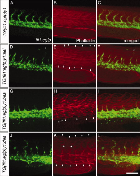
Fig. 4 ISVs follow the irregular somite boundaries in Notch mutants. Wildtype (A-C) and Notch mutant embryos, aei (D–F), bea (G-I), and des (J-L), were stained with phalloidin to reveal somitic actin fibres (in red; B,E,H,K). The ISV vascular pattern revealed by the green fluorescence of the fli1:EGFP embryos (A,D,G,J) is seen correlating with the somitic boundaries (B,E,H,K) in the merged images (C,F,I,L). Asterisks indicate misplaced ISV over somite. Scale bar = 100 μm.
Acknowledgments
This image is the copyrighted work of the attributed author or publisher, and
ZFIN has permission only to display this image to its users.
Additional permissions should be obtained from the applicable author or publisher of the image.
Full text @ Dev. Dyn.