
- Title
-
In vivo imaging of zebrafish retinal cells using fluorescent coumarin derivatives
- Authors
- Watanabe, K., Nishimura, Y., Oka, T., Nomoto, T., Kon, T., Shintou, T., Hirano, M., Shimada, Y., Umemoto, N., Kuroyanagi, J., Wang, Z., Zhang, Z., Nishimura, N., Miyazaki, T., Imamura, T., and Tanaka, T.
- Source
- Full text @ BMC Neurosci.
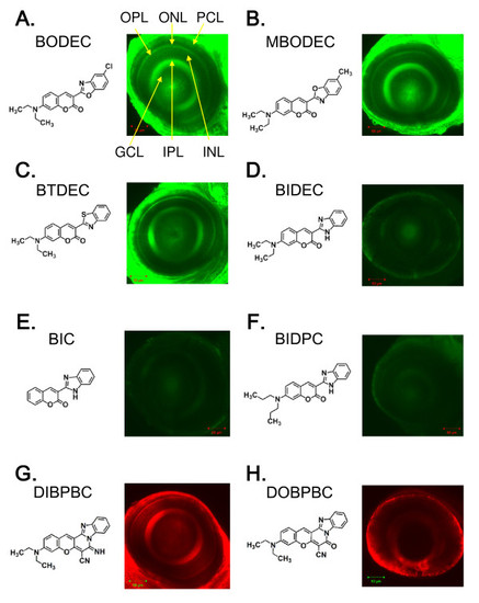
Identification of coumarin derivatives visualizing the multiple layers of the zebrafish retina in vivo. Zebrafish larvae at 6 dpf were stained with BODEC (A), MBODEC (B), BTDEC (C), BIDEC (D), BIC (E), BIDPC (F), DIBPBC (G) and DOBPBC (H). The retinas were visualized by confocal laser scanning microscopy. The zebrafish retinas are clearly visualized by BODEC (A), MBODEC (B), BTDEC (C) and DIBPBC (G). OPL: outer plexiform layer; ONL: outer nuclear layer; PCL: photoreceptor layer; GCL: ganglion cell layer; IPL: inner plexiform layer; INL: inner nuclear layer. |
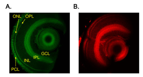
The coumarin dyes are fixable. Zebrafish larvae at 6 dpf were stained with BODEC (A) or DIBPBC (B) and fixed in 4% paraformaldehyde. The retinas were sectioned and visualized by confocal laser scanning microscopy. The zebrafish retinas are clearly visualized similar to the in vivo imaging. |
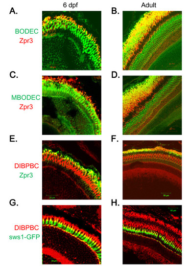
Staining of rod and UV-sensitive cone photoreceptor cells with the coumarin derivatives. Retinal sections from wild-type AB larvae (A, C and E) and adults (B, D and F) were labeled with zpr3, followed by labeling with secondary antibodies conjugated with Alexa fluorophores and counter-staining with BODEC (A and B), MBODEC (C and D) or DIBPBC (E and F). The fluorescent signals of the coumarin derivatives (green in A-D; red in E and F) partly overlap (yellow in A-F) with those of zpr3 (red in A-D; green in E and F). Retinal sections from a Tg (sws1:GFP) larva (G) and adult (H) were counter-stained with DIBPBC. The fluorescent signals of DIBPBC (red) partly overlap (yellow) with those of zpr3 (green). |
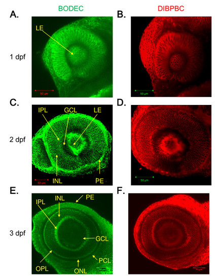
In vivo imaging of zebrafish retinal development using the coumarin derivatives. Zebrafish larvae at 1 dpf (A and B), 2 dpf (C and D) and 3 dpf (E and F) were stained with BODEC (A, C and E) or DIBPBC (B, D and F). After the staining, the retinas were visualized by confocal laser scanning microscopy. The development of the retinal structures is clearly visualized. |
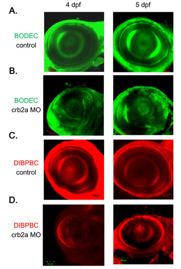
In vivo imaging of the zebrafish retina in a genetic model of retinopathy using the coumarin derivatives. Control zebrafish (A and C) and crb2a morphants (B and D) at 4 and 5 dpf were stained with BODEC (A and B) or DIBPBC (C and D). The retinas were visualized by confocal laser scanning microscopy. The retinal disorganization in the crb2a morphants is clearly visualized by both BODEC and DIBPBC. |
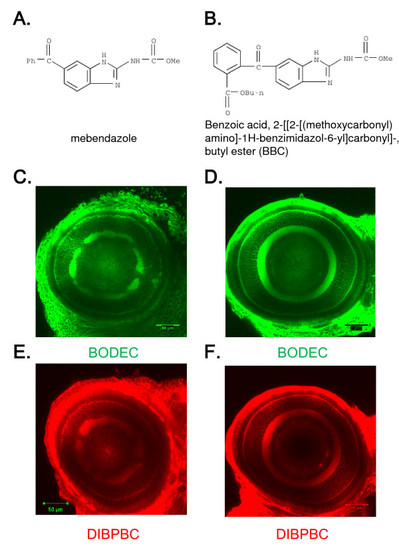
In vivo imaging of the zebrafish retina treated with toxic chemicals. Zebrafish larvae were treated with mebendazole (A) or BBC (B) from 1 to 6 dpf. The larvae were stained with BODEC (C and D) or DIBPBC (E and F). The retinas were visualized by confocal laser scanning microscopy. Both BODEC and DIBPBC clearly visualize the disorganization of the IPL in the zebrafish treated with mebendazole (C and E) but not with BBC (D and F). |
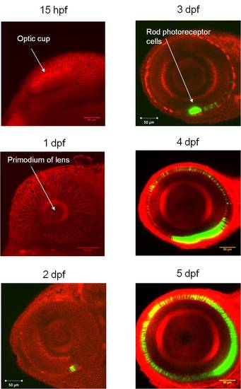
In vivo imaging of the zebrafish retina by combining the coumarin derivatives and transgenic zebrafish expressing GFP in rod photoreceptor cells. Tg(rh:GFP) zebrafish from 1 to 5 dpf were stained with DIBPBC. The retinas were visualized by confocal laser scanning microscopy. The development of rod photoreceptor cells is visualized with high resolution by the counter-staining with DIBPBC. |
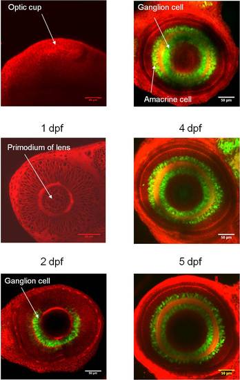
In vivo imaging of the zebrafish retina by combining the coumarin derivatives and transgenic zebrafish expressing Kaede in retinal ganglion cells and amacrine cells. Tg(huc:Kaede) zebrafish from 1 to 5 dpf were stained with DIBPBC. The retinas were visualized by confocal laser scanning microscopy. The development of retinal ganglion cells and amacrine cells is visualized with high resolution by the counter-staining with DIBPBC. |