
FIGURE
Fig. 5
- ID
- ZDB-FIG-101013-13
- Publication
- Watanabe et al., 2010 - In vivo imaging of zebrafish retinal cells using fluorescent coumarin derivatives
- Other Figures
- All Figure Page
- Back to All Figure Page
Fig. 5
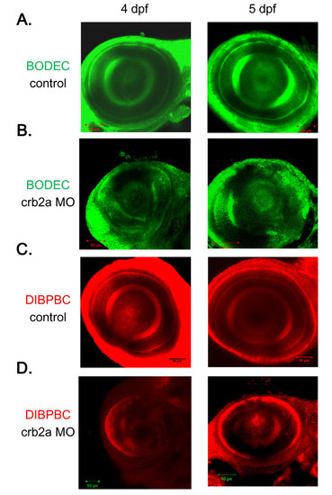
In vivo imaging of the zebrafish retina in a genetic model of retinopathy using the coumarin derivatives. Control zebrafish (A and C) and crb2a morphants (B and D) at 4 and 5 dpf were stained with BODEC (A and B) or DIBPBC (C and D). The retinas were visualized by confocal laser scanning microscopy. The retinal disorganization in the crb2a morphants is clearly visualized by both BODEC and DIBPBC. |
Expression Data
Expression Detail
Antibody Labeling
Phenotype Data
Phenotype Detail
Acknowledgments
This image is the copyrighted work of the attributed author or publisher, and
ZFIN has permission only to display this image to its users.
Additional permissions should be obtained from the applicable author or publisher of the image.
Full text @ BMC Neurosci.