
IMAGE
Fig. 1
- ID
- ZDB-IMAGE-101013-9
- Publication
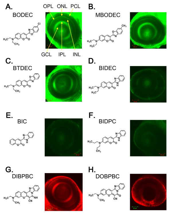
- Watanabe et al., 2010 - In vivo imaging of zebrafish retinal cells using fluorescent coumarin derivatives
- All Figures
- Figures for Watanabe et al., 2010
Image
|
Figure Caption
Fig. 1 Identification of coumarin derivatives visualizing the multiple layers of the zebrafish retina in vivo. Zebrafish larvae at 6 dpf were stained with BODEC (A), MBODEC (B), BTDEC (C), BIDEC (D), BIC (E), BIDPC (F), DIBPBC (G) and DOBPBC (H). The retinas were visualized by confocal laser scanning microscopy. The zebrafish retinas are clearly visualized by BODEC (A), MBODEC (B), BTDEC (C) and DIBPBC (G). OPL: outer plexiform layer; ONL: outer nuclear layer; PCL: photoreceptor layer; GCL: ganglion cell layer; IPL: inner plexiform layer; INL: inner nuclear layer.
Acknowledgments
This image is the copyrighted work of the attributed author or publisher, and
ZFIN has permission only to display this image to its users.
Additional permissions should be obtained from the applicable author or publisher of the image.
Full text @ BMC Neurosci.