
Fig. 3
- ID
- ZDB-FIG-101013-11
- Publication
- Watanabe et al., 2010 - In vivo imaging of zebrafish retinal cells using fluorescent coumarin derivatives
- Other Figures
- All Figure Page
- Back to All Figure Page
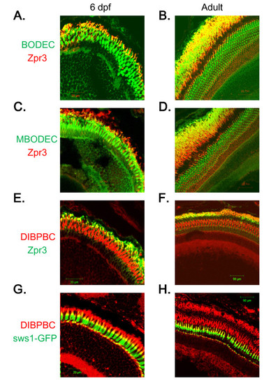
Staining of rod and UV-sensitive cone photoreceptor cells with the coumarin derivatives. Retinal sections from wild-type AB larvae (A, C and E) and adults (B, D and F) were labeled with zpr3, followed by labeling with secondary antibodies conjugated with Alexa fluorophores and counter-staining with BODEC (A and B), MBODEC (C and D) or DIBPBC (E and F). The fluorescent signals of the coumarin derivatives (green in A-D; red in E and F) partly overlap (yellow in A-F) with those of zpr3 (red in A-D; green in E and F). Retinal sections from a Tg (sws1:GFP) larva (G) and adult (H) were counter-stained with DIBPBC. The fluorescent signals of DIBPBC (red) partly overlap (yellow) with those of zpr3 (green). |