
IMAGE
Fig. 4
- ID
- ZDB-IMAGE-101013-12
- Publication
- Watanabe et al., 2010 - In vivo imaging of zebrafish retinal cells using fluorescent coumarin derivatives
- All Figures
- Figures for Watanabe et al., 2010
Image
|
Figure Caption
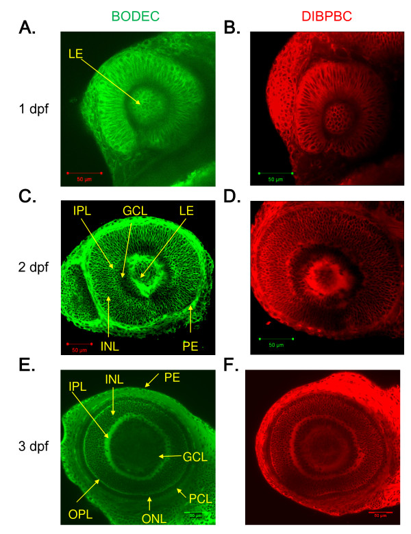
Fig. 4 In vivo imaging of zebrafish retinal development using the coumarin derivatives. Zebrafish larvae at 1 dpf (A and B), 2 dpf (C and D) and 3 dpf (E and F) were stained with BODEC (A, C and E) or DIBPBC (B, D and F). After the staining, the retinas were visualized by confocal laser scanning microscopy. The development of the retinal structures is clearly visualized.
Acknowledgments
This image is the copyrighted work of the attributed author or publisher, and
ZFIN has permission only to display this image to its users.
Additional permissions should be obtained from the applicable author or publisher of the image.
Full text @ BMC Neurosci.