
- Title
-
Generation of oocyte-specifically expressed cre transgenic zebrafish for female germline excision of loxp-flanked transgene
- Authors
- Liu, X., Li, Z., Emelyanov, A., Parinov, S., and Gong, Z.
- Source
- Full text @ Dev. Dyn.

Transient expression of pKRT8-LGLR in zebrafish embryos by co-injection of pCMV-CRE or pZP3-CRE. A: Schematic representation of chimeric DNA constructs, pKRT8-LGLR, pCMV-CRE, and pZP3-CRE. B: A 48 hours postfertilization (hpf) embryo injected with pKRT8-LGLR only, showing only GFP expression. C,D: A same 48 hpf embryo co-injection of pKRT8-LGLR and pCMV-CRE, showing both green fluorescent protein (GFP) expression under a blue light (C) and red fluorescent protein (RFP) expression under a yellow light (D). E,F: A same 48 hpf embryo co-injection of pKRT8-LGLR and pZP3-CRE, showing both GFP (E) and RFP (F) expression. Scale bars = 500 μm. |

Co-existence of red fluorescent protein (RFP) expression and cre DNA in Tg(zp3:cre, krt8:rfp) F1 embryos. A: Skin-specific RFP expression in Tg(zp3:cre, krt8:rfp) embryos: 24 hours postfertilization (hpf; upper) and 72 hpf (lower). B: Presence of cre DNA in RFP-positive embryos. Genomic DNAs were prepared from 5 days postfertilization embryos individually and cre DNA was detected by polymerase chain reaction (PCR). The picture shows nine RFP-positive embryos and one RNA-negative embryo. P, positive PCR control with pZP3-CRE template; N, negative PCR control without template; M, 100-bp DNA marker (Promega). C: Demonstration of the Cre-mediated excision of floxed egfp in Tg(zp3:cre, krt8:rfp) transgenic fish. PCR was performed for both RFP-positive and -negative embryos using primers P1 and R1 (upper) as indicated in the diagram with expected sizes of PCR products before and after Cre-mediated excision. Presence of cre DNA was also amplified by PCR using a pair of cre-specific primers (lower). The 850-bp recombined fragment and cre DNA are co-existed in skin RFP expressing embryos (RFP+) but not in the non-RNA expressing embryos (RFP-). Primers P1+G1 amplified 750-bp unexcised gfp fragment. P, positive PCR control with templates pKRT8-LGLR (upper) or pZP3-CRE (lower); N, negative PCR control without template; M, 1-kb DNA marker. In the diagram, LoxP, EGFP, and RFP DNAs are indicated by blue triangles, green and red boxes, respectively. Scale bars = 500 μm. |

Cre RNA expression in Tg(zp3:cre, krt8:rfp) transgenic zebrafish. A,B: cre RNA expression in 2-72 hours postfertilization (hpf) embryos (A) and in adult transgenic fish (B) as detected by reverse transcriptase-polymerase chain reaction (RT-PCR). Embryonic stages in hpf (A) and tissue sources (B) are indicated at the top of each lane. Whole-male transgenic fish (WFB) and selected tissues from female transgenic fish were used for RNA preparation. P, positive control with pZP3-CRE plasmid; N, negative control without template. β-actin probes were used for RT-PCR control. C,D: Ovary expression of cre mRNA in Tg(zp3:cre, krt8:rfp) transgenic female (C) and zp3 mRNA wild-type female (D). Whole-mount in situ hybridization detection was performed with isolated ovary tissues and both cre and zp3 mRNAs were detected in developing oocytes (arrows) but not in the mature oocyte. EXPRESSION / LABELING:
|
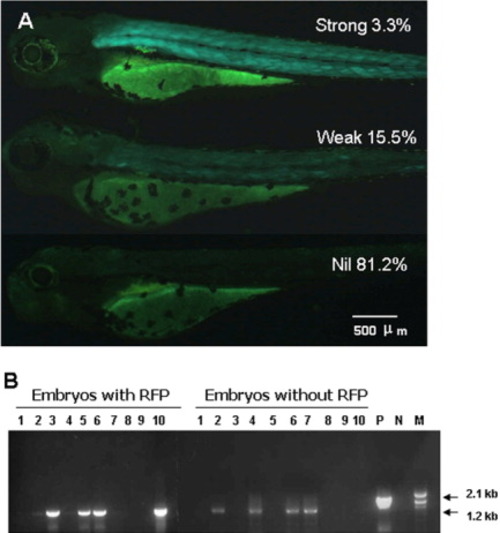
Test of Cre-mediated recombination at the chromosome level by breeding of Tg(zp3:cre, krt8:rfp) and Tg(mylz2:loxP-EGFP-loxP)gz3. A: The 48 hpf embryos with strong, weak, and nil green fluorescent protein (GFP) expression from a cross between female Tg(zp3:cre, krt8:rfp) and male Tg(mylz2:loxP-EGFP-loxP)gz3. Percentages of each type of embryos based on GFP expression are indicated. B: Polymerase chain reaction (PCR) detection of Cre-mediated recombination in offspring of female double transgenic fish Tg(zp3:cre, krt8:rfp)/Tg(mylz2:loxP-EGFP-loxP)gz3 and male wild-type fish. In both skin RFP-expressing (cre transgenic) and non-RFP-expressing (cre non-transgenic) embryos (4 days postfertilization), approximately half of them showed Cre-mediated recombination as anticipated from the female germline excision while the other half were loxP non-transgenic individuals. The expected PCR products are 2.1 kb and 1.2 kb, respectively, before and after the Cre-mediated recombination. P, positive PCR control with pMYLZ2-loxP-EGFP-loxP plasmid that is used to generate Tg(mylz2:loxP-EGFP-loxP)gz3; N, negative PCR control without a template DNA; M, 1-kb DNA ladder. Scale bar = 500 μm. EXPRESSION / LABELING:
|

Activation of loxP-blocked reporter gene expression. A,B: A 48 hours postfertilization (hpf) embryo from a cross between a female Tg(zp3:cre, krt8:rfp) and male Tg(EF:loxP-mCherry-loxP-egfp) to show mCherry (A) and enhanced green fluorescent protein (EGFP; B) expression. C,D: A 48-hpf embryo from a cross between a female wild-type fish and male Tg(EF:loxP-mCherry-loxP-egfp) to show mCherry (C) and GFP (D) expression. E,F: View of mCherry (E) and GFP (F) expression in the ovary from a 1.5-month-old female double transgenic fish Tg(Zp3:Cre, Krt8:RFP)/ Tg(EF:loxp-mCherry-loxp-egfp. G,H: View of mCherry (G) and GFP (H) expression in the ovary from a 1.5-month-old female Tg(EF:loxp-mCherry-loxp-egfp fish. Scale bars = 100 μm. |