- Title
-
Live analysis of endodermal layer formation identifies random walk as a novel gastrulation movement
- Authors
- Pézeron, G., Mourrain, P., Courty, S., Ghislain, J., Becker, T.S., Rosa, F.M., and David, N.B.
- Source
- Full text @ Curr. Biol.
|
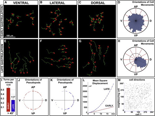
Endodermal Cells Disperse with a Random Walk Movement during Early Gastrulation (A–C and E–G) Representative examples of 30 min tracks with 1 min intervals of endodermal cells in Et(CLG-YFP)smb602 embryos during early (55%–70% epiboly [A–C]) or late (75%–90% epiboly [E–G]) gastrulation (similar tracks were obtained for more than 400 cells on about five embryos per position and stage). During early gastrulation cells move in a nonoriented fashion, whereas during late gastrulation they undergo convergence-extension movements (i.e., dorsal cells migrate anteriorly, lateral cells converge toward the embryonic axis, and ventral cells migrate toward the vegetal pole). Red arrowheads indicate the direction of the last tracked movement. The animal pole is to the top, and for lateral views, dorsal is to the right. (D and H) Rose diagrams representing the directions of endodermal cell movements. During early gastrulation cells migrate in all directions (D) compared to the oriented migration of converging cells during late gastrulation (H). Early gastrulation data were obtained from four time lapses on lateral views (D) and late gastrulation data from four time-lapses on lateral views (H). (I) The graph indicates the average number of turns per minute per cell during early gastrulation (n = 164 cells) or during late gastrulation (n = 68 cells). On average, cells maintained the same direction (angle of turn < 45°) for only 2.21 min compared to 4.95 min for converging cells (p < 0.0001). Error bars represent standard errors. (J and K) Polar plots of the distribution of the outgrowth positions of pseudopods relative to the cell center for lateral cells during early (J) and late (K) gastrulation. Each pseudopod was counted only once, even though pseudopods often persisted for more than one frame. For each diagram, 20 cells from four embryos were analyzed over a 30 min period. (L) Plot (dot and square) and curve fit (line) of the MSD of cells during early (red) and late (blue) gastrulation, showing that whereas converging cells have an oriented migration (parabolic fit, R = 0.999, n = 164 cells), cells move in a random walk during early gastrulation (linear fit, R = 0.998, n = 58 cells). (M) Scatter plot of the direction of a cell and of its closest neighbor, showing that cell movements are not coordinated (R = 0.12, n = 589). |
|
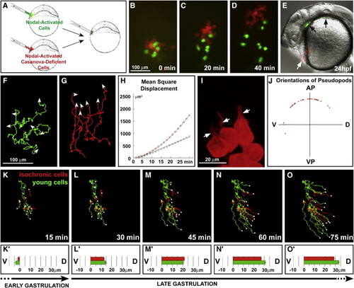
Control of Random Walk Behavior (A–J) Random walk is inducible by Nodal and depends on casanova. In (A), a schematic of the experimental procedure is illustrated. (B–D) Nodal-activated cells (green, Tar* cells) and Nodal-activated cells coinjected with a morpholino against casanova (red, Tar*-MOcasanova cells) were transplanted into wild-type host embryos and monitored during early gastrulation. (E) At 24 hpf, Tar* cells are found within the endoderm (pharynx, black arrows), whereas Tar*-MOcasanova cells contribute to the hatching gland (white arrow). (F and G) Shown are representive examples of 40 min tracks with 1 min intervals of Tar* and Tar*-MOcasanova cells after transplantation. (H) A MSD plot reveals that Tar* cells (green) migrate in a random walk (linear fit, R = 0.996), whereas Tar*-MOcasanova cells (red) display an oriented migration (parabolic fit, R = 0.999). For each population, 30 cells from four embryos were analyzed. (I) Shown is a representive example of Tar*-MOcasanova cell morphology during early gastrulation. Cells develop large cytoplasmic processes (white arrows). In (J), a polar plot illustrates the distribution of the outgrowth positions of pseudopods relative to the cell center of Tar*-MOcasanova cells during early gastrulation. Seventeen cells from three embryos were analyzed over 20 min. (K–O′) Transition to a convergence movement is induced by the embryonic environment. Nodal-activated cells from midblastula (4 hpf) embryos (young cells, green) and late-blastula (5 hpf) embryos (isochronic cells, red) were transplanted into late-blastula (5 hpf) host embryos. Shown is a representative example of four independent experiments. (K–O) Shown are tracks of both cell populations after 15, 30, 45, 60, or 75 min of monitoring. Host midgastrulation (70% epiboly) corresponds to t = 15 min. The white dots indicate the end position of each track. (K′)–(O′) illustrates the mean net displacement toward the dorsal side for each 15 min interval. During host early gastrul2870% epiboly) corresponds to t = 15 min. The white dots indicate the end position of each track. (K′)–(O ion, both cell populations first migrate randomly without any dorsal bias (K′). They simultaneously start to converge dors |
|
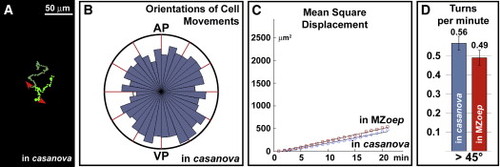
Random Walk Does Not Depend on Cell Interactions One or a few Nodal-activated cells were transplanted into endoderm-deficient casanova morphant embryos and into endoderm- and dorsal mesoderm-deficient MZoep mutant embryos. (A) Representative example of 50 min tracks (with 1 min intervals) of two cells derived from one cell transplanted into a casanova morphant embryo. (B) Rose diagram of the directions of cell movements shows that in casanova morphant, transplanted cells migrate in all directions (n = 11 cells from three embryos). (C) Plot (dot) and curve fit (line) of the MSD showing that these cells move in a random walk during early gastrulation (in casanova: linear fit, R = 0.992, n = 11 cells from three embryos; in MZoep: linear fit, R = 0.997, n = 14 cell from four embryos). (D) The graph illustrates the average number of turns per minute per Nodal-activated cell. Even in the absence of other endodermal cells (in casanova) or of hypoblastic cells (in MZoep), Nodal-activated cells frequently change their direction. Error bars represent standard errors. |
|
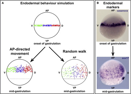
Random Walk Is Sufficient for Endodermal Cells to Colonize the Yolk Surface (A) Cell migration was mathematically simulated to test how the observed pattern of endodermal cells at midgastrulation can be achieved. At the beginning of gastrulation, 100 cells were localized at the margin of the blastoderm (50% epiboly). Depending on their position along the dorsoventral axis, cells were marked in red (dorsal), blue (lateral), or green (ventral). An oriented motion toward the animal pole cannot account for the pattern observed in vivo at 75% epiboly. In contrast, a random walk efficiently spreads cells over the yolk, with the expansion of the layer being limited both by cell speed and by the margin. At midgastrulation (75% epiboly), cells have reached a position similar to the expression pattern of endodermal markers and have partially mixed. (B) Shown is the distribution of endodermal cells at the onset of gastrulation and at midgastrulation as assayed by sox17 expression. EXPRESSION / LABELING:
|
|
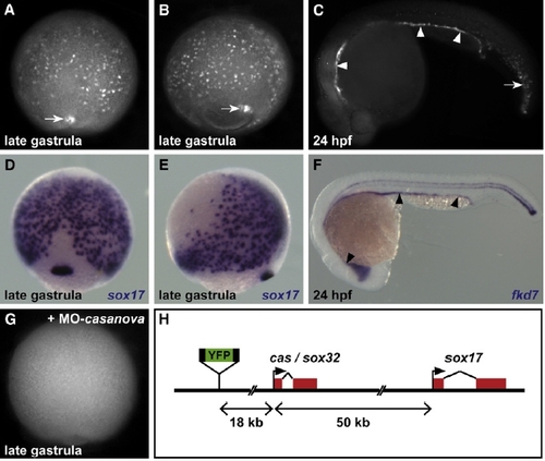
The Et(CLG-YFP)smb602 Line Specifically Expresses YFP in Endodermal Cells (A and B) Live imaging of Et(CLG-YFP)smb602 embryos reveals YFP expression in isolated hypoblastic cells located on the yolk surface and in the forerunner cells (white arrows) during gastrulation. (C) At 20 hpf, YFP is detected in all endodermal derivatives (arrowheads) and in derivatives of the forerunner cells (arrow). (D–F) Location of endodermal cells during gastrulation (D and E) and of endodermal derivatives at 24 hpf ([F], arrowheads) revealed by the expression of the endodermal markers sox17 and fkd7. (G) YFP expression is abolished in Et(CLG-YFP)smb602 embryos injected with casanova-Morpholino. (H) In the Et(CLG-YFP)smb602 line, the CLGY reporter is integrated upstream of the casanova and sox17 genomic loci. (A, D, and G) A dorsal view is presented; the animal pole is to the top. (B and E) A lateral view is presented; the animal pole is to the top and dorsal is to the right. (C and F) A lateral view is presented; anterior is to the left. EXPRESSION / LABELING:
|
|
Endodermal Cells Do Not Behave Like Mesodermal Cells during Early Gastrulation (A–C) Shown is the evolution of a clone of mesodermal cells (labeled in red) transplanted at the margin of an Et(CLG-YFP)smb602 embryo (in green) from 60% to 70% epiboly (lateral view, animal pole to the top, dorsal to the right). (D) Net path of internalized mesodermal cells (red) and endodermal cells (green) from time lapse in (A)–(C) is shown. Whereas mesodermal cells move toward the animal pole, endodermal cells do not show any preferred direction. |

