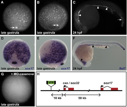
The Et(CLG-YFP)smb602 Line Specifically Expresses YFP in Endodermal Cells (A and B) Live imaging of Et(CLG-YFP)smb602 embryos reveals YFP expression in isolated hypoblastic cells located on the yolk surface and in the forerunner cells (white arrows) during gastrulation. (C) At 20 hpf, YFP is detected in all endodermal derivatives (arrowheads) and in derivatives of the forerunner cells (arrow). (D–F) Location of endodermal cells during gastrulation (D and E) and of endodermal derivatives at 24 hpf ([F], arrowheads) revealed by the expression of the endodermal markers sox17 and fkd7. (G) YFP expression is abolished in Et(CLG-YFP)smb602 embryos injected with casanova-Morpholino. (H) In the Et(CLG-YFP)smb602 line, the CLGY reporter is integrated upstream of the casanova and sox17 genomic loci. (A, D, and G) A dorsal view is presented; the animal pole is to the top. (B and E) A lateral view is presented; the animal pole is to the top and dorsal is to the right. (C and F) A lateral view is presented; anterior is to the left.
|

