
- Title
-
cadherin-6 Message expression in the nervous system of developing zebrafish
- Authors
- Liu, Q., Liu, B., Wilson, A.L., and Rostedt, J.
- Source
- Full text @ Dev. Dyn.
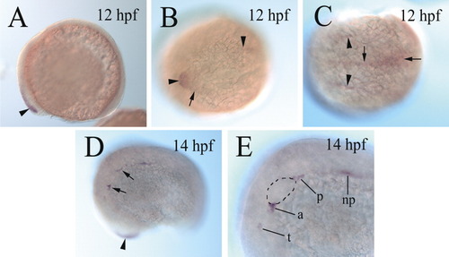
cadherin-6 expression in 12 and 14 hours postfertilization (hpf) zebrafish embryos. A: A lateral view of a whole-mount embryo, showing cadherin-6 expression (arrowhead) in the anterodorsal neural keel. B,C: Dorsal views of a whole-mount embryo (anterior to the left). B: The horizontal arrowhead indicates the anterodorsal cadherin-6 expression, whereas the vertical arrowhead points to the anterior limit of the cadherin-6 expression at the midline. The arrow indicates the lateral edge of the expression domain. C: The arrowheads point to cadherin-6 expression at the edge of the neural keel, whereas the vertical arrow indicates the cadherin-6 expression domain at the midline. cadherin-6 expression near the posterior end of the neural keel is indicated by the horizontal arrow. D: The arrowhead indicates the cadherin-6 expression domain in the head region (out of focus). The two arrows point to two of the cadherin-6-expressing regions. E: A higher magnification view of the labeled regions indicated by the arrows in D. The otic placode (op) is outlined with dashed lines. a, anterior lateral line placode area; np, nephric duct; p, posterolateral line placode/ganglion; t, trigeminal placode/ganglion. EXPRESSION / LABELING:
|
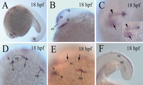
cadherin-6 expression in 18 hours postfertilization (hpf) embryos. A,B: B is a higher magnification image of the head region of the embryo in A, with anterior to the left and dorsal up. C: A higher magnification image of a dorsolateral view of the forebrain (anterior to the left), focusing on the cadherin-6 expression domain (arrowhead) in the dorsal forebrain, whereas the inset shows the same brain, focusing on cadherin-6 expression domain (arrow) in the ventral diencephalon. D: A higher magnification of the hindbrain region (anterior to the left and dorsal up) of the embryo in A. E: A higher magnification of a dorsolateral view of the anterior spinal cord (sc) region (with anterior to the left) of the embryo in A. The two arrows indicate cadherin-6 expression in the dorsal spinal cord. F: A lateral view (anterior to the left and dorsal up) of the tail region of an embryo showing cadherin-6 expression in the dorsal spinal cord. The otic placode in D is outlined with dashed lines. di, diencephalon; e, epiphysis; h, hindbrain; nc, notochord; or, optic recess. The remaining abbreviations are the same as in Figure 1. EXPRESSION / LABELING:
|
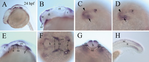
Expression of cadherin-6 message in 24 hours postfertilization (hpf) embryos. A,B: B is a higher magnification of the fore- and midbrain regions of the embryo in A, with anterior to the left and dorsal up. C,D: Higher magnification dorsolateral views (anterior to the left) of the forebrain region of the same whole-mount embryo, with C focusing on cadherin-6 expression in the dorsal forebrain (arrowhead) and D focusing on cadherin-6 expression in the ventral diencephalon (arrow). E: A higher magnification of the hindbrain region of the embryo in A (anterior to the left and dorsal up). F: A higher magnification dorsal view (anterior to the left) of the hindbrain region of a whole-mount embryo. The asterisk indicates cadherin-6 expression outside the nervous tissue. G: A frontal view of the hindbrain region (dorsal up) at the level of otic vesicle (ov), of a whole-mount embryo. H: A higher magnification lateral view (anterior to the left and dorsal up) of a whole-mount tail region. c, cerebellum; sa, statoacoustic ganglion. The remaining abbreviations are the same as in Figures 1 and 2. EXPRESSION / LABELING:
|
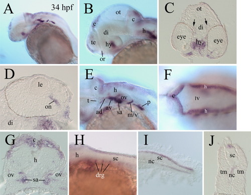
cadherin-6 expression in 34 hours postfertilization (hpf) embryos. A: A lateral view (anterior to the left and dorsal up) of a whole-mount embryo. B,E: Higher magnifications of the anterior half and posterior half, respectively, of the embryo in A. C,D,G,J: Cross-sections of embryos processed for whole-mount in situ hybridization. C (dorsal up) shows a frontal view of the cadherin-6 expression in the hypothalamus (hy) and the pretectal area (arrows). D: A higher magnification of an eye section (dorsal to the left). F: A dorsal view of the hindbrain region (anterior to the left). G: A cross-section (dorsal up) through the cerebellum and anterior hindbrain region. H,I Lateral views of the anterior spinal cord and tail regions (anterior to the left and dorsal up), respectively, whole-mount embryos. J: A cross-section (dorsal up) through the midtrunk region. ad, anterodorsal lateral line ganglion; av, anteroventral lateral line ganglion; IV, the fourth ventricle; le, lens; m/v, medial lateral line ganglion and vagal ganglion; tm, trunk muscles. The remaining abbreviations are the same as in the previous figures. EXPRESSION / LABELING:
|
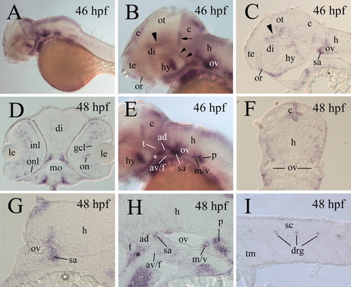
cadherin-6 expression in 46-48 hpf embryos. A,B,E: Lateral views of whole-mount embryos (anterior to the left and dorsal up), whereas the remaining panels are sections from whole-mount embryos processed for in situ hybridization. C,H,J: Parasagittal sections (anterior to the left and dorsal up). D,F,G: Cross-sections (dorsal up). B,E: Higher magnification images of the anterior half and posterior half, respectively, of the head region of the embryo in A. B: The large arrowhead and two smaller arrowheads point to the cadherin-6 expression domains in the dorsal thalamic and the posterior hypothalamic regions, respectively, whereas the arrow indicates cadherin-6 expression in the anterior cerebellum. E: The asterisk indicates cadherin-6 expression outside the nervous system. C: The arrowhead indicates the dorsal thalamic cadherin-6 expression. G: A higher magnification of the hindbrain at the level of the otic vesicle. H: A higher magnification image of the hindbrain, with the asterisk indicating cadherin-6 expression outside the nervous system. av/f, anteroventral lateral line and facial ganglia; gcl, ganglion cell layer; inl, inner nuclear layer; onl; outer nuclear layer. Other abbreviations are the same as in the previous figures. EXPRESSION / LABELING:
|