
|
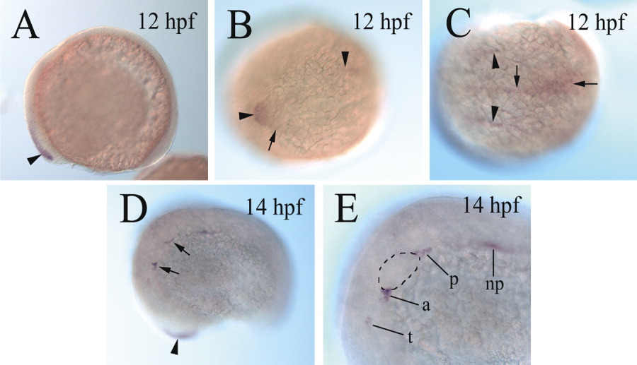
Fig. 1 cadherin-6 expression in 12 and 14 hours postfertilization (hpf) zebrafish embryos. A: A lateral view of a whole-mount embryo, showing cadherin-6 expression (arrowhead) in the anterodorsal neural keel. B,C: Dorsal views of a whole-mount embryo (anterior to the left). B: The horizontal arrowhead indicates the anterodorsal cadherin-6 expression, whereas the vertical arrowhead points to the anterior limit of the cadherin-6 expression at the midline. The arrow indicates the lateral edge of the expression domain. C: The arrowheads point to cadherin-6 expression at the edge of the neural keel, whereas the vertical arrow indicates the cadherin-6 expression domain at the midline. cadherin-6 expression near the posterior end of the neural keel is indicated by the horizontal arrow. D: The arrowhead indicates the cadherin-6 expression domain in the head region (out of focus). The two arrows point to two of the cadherin-6-expressing regions. E: A higher magnification view of the labeled regions indicated by the arrows in D. The otic placode (op) is outlined with dashed lines. a, anterior lateral line placode area; np, nephric duct; p, posterolateral line placode/ganglion; t, trigeminal placode/ganglion.