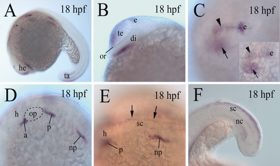
Fig. 2 cadherin-6 expression in 18 hours postfertilization (hpf) embryos. A,B: B is a higher magnification image of the head region of the embryo in A, with anterior to the left and dorsal up. C: A higher magnification image of a dorsolateral view of the forebrain (anterior to the left), focusing on the cadherin-6 expression domain (arrowhead) in the dorsal forebrain, whereas the inset shows the same brain, focusing on cadherin-6 expression domain (arrow) in the ventral diencephalon. D: A higher magnification of the hindbrain region (anterior to the left and dorsal up) of the embryo in A. E: A higher magnification of a dorsolateral view of the anterior spinal cord (sc) region (with anterior to the left) of the embryo in A. The two arrows indicate cadherin-6 expression in the dorsal spinal cord. F: A lateral view (anterior to the left and dorsal up) of the tail region of an embryo showing cadherin-6 expression in the dorsal spinal cord. The otic placode in D is outlined with dashed lines. di, diencephalon; e, epiphysis; h, hindbrain; nc, notochord; or, optic recess. The remaining abbreviations are the same as in Figure 1.
Image
Figure Caption
Figure Data
Acknowledgments
This image is the copyrighted work of the attributed author or publisher, and
ZFIN has permission only to display this image to its users.
Additional permissions should be obtained from the applicable author or publisher of the image.
Full text @ Dev. Dyn.

