
- Title
-
Cxcl8b and Cxcr2 Regulate Neutrophil Migration through Bloodstream in Zebrafish
- Authors
- Zuñiga-Traslaviña, C., Bravo, K., Reyes, A.E., Feijóo, C.G.
- Source
- Full text @ J Immunol Res
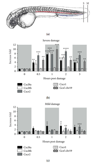
Severe and mild damage differentially regulate the transcription of cxcl8 paralogues in zebrafish. (a) Diagram showing the location of severe (S) and mild (M) damage on the caudal region and caudal fin of the embryo, respectively. The red line corresponds to the caudal artery, and the blue line to the caudal vein. (b, c) Transcription levels of cxcl8a, cxcl8b, cxcr2, cxcr1, and gcsf-chr19 were quantified by qPCR after (b) severe or (c) mild damage. Data are presented as fold of change over each level at 0 hours post damage and normalized to b-actin1. * p value < 0.05; ** p value < 0.01; *** p value < 0.005. |

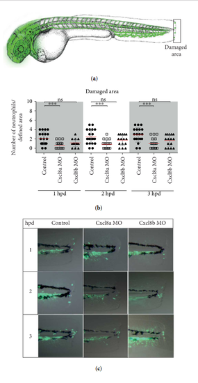
Neutrophil migration decreases when Cxcl8a or Cxcl8b are inhibited during severe damage. (a) Diagram showing the quantified neutrophils in two areas. (b) Lateral view of the caudal section of the embryo tail at 1, 2, and 3 hpd in control and morphant embryos. (c) Quantified neutrophils at the damaged area at 1, 2, and 3 hpd. (d) Quantified neutrophils in circulation during homeostasis, after severe damage and in the absence of Cxcl8a or Cxcl8b function. The neutrophils were quantified at 1, 2, and 3 hpd. value * p < 0.05; ** p value < 0.01; *** p value < 0.001; **** p value < 0.0001. PHENOTYPE:
|
Neutrophil migration decreases when Cxcl8a, but not Cxcl8b, is inhibited during mild damage. (a) Diagram showing the quantified neutrophils at the wound area. (b) Quantified neutrophils at damaged area at 1, 2, and 3 hpd. (c) Lateral view of the caudal section of the embryo tail at 1, 2, and 3 hpd in the control and morphant embryos. value *** p < 0.005. PHENOTYPE:
|
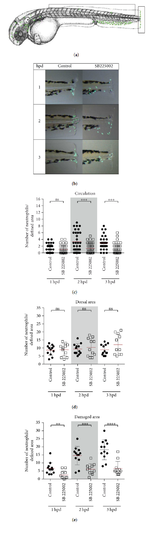
Cxcr2 inhibition decreases neutrophil entrance to the bloodstream and tissue infiltration in severe damage. (a) Diagram showing the quantified neutrophils, in circulation and at the dorsal and damaged area. (b) Neutrophils in circulation at 1, 2, and 3 hpd in control embryos and treated with the inhibitor SB225002. (c) Lateral view of the caudal section of the embryo tail at 1, 2, and 3 hpd in the control and treated embryos. (d, e) Quantified neutrophils at the dorsal and damaged areas at 1, 2, and 3 hpd. value ** p < 0.01; *** p value < 0.005; **** p value < 0.0001. |
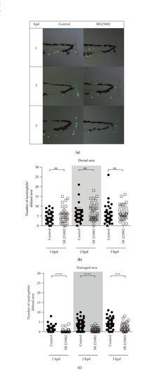
Cxcr2 inhibition decreases neutrophil infiltration in mild damage. (a) Lateral view of the caudal section of the embryo tail at 1, 2, and 3 hpd in the control and treated embryos. Demarked with a white rectangle are the two quantified areas. (b, c) Quantified neutrophils at the dorsal and damaged areas at 1, 2, and 3 hpd. *** p value < 0.005; **** p value < 0.0001. |

Morphant phenotype. (A, B, C) Representative images of the different phenotypes observed after Cxcl8a or Cxl8b morpholino microinjection. (A) Control phenotype showing vasculature disruption. (B) Morphant phenotype used in the damage assays, showing 2-3 disruptions only in intersegmental vessels (arrow). (C) Severe phenotype, with the vasculature showing more than three alterations. PHENOTYPE:
|