
- Title
-
BDNF Expression in Larval and Adult Zebrafish Brain: Distribution and Cell Identification
- Authors
- Cacialli, P., Gueguen, M.M., Coumailleau, P., D'Angelo, L., Kah, O., Lucini, C., Pellegrini, E.
- Source
- Full text @ PLoS One
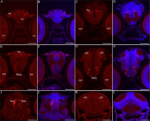
Bdnf mRNA are expressed in the brain of 7 days old zebrafish. Olfactory rosettes (A-B), telencephalon (A-F), preoptic area (E-H), dorsal thalamus, (I-J), optic tectum (G-H and K-L). Figures B, D, F, H, J and L show cell nuclei labeled with DAPI. OR: olfactory rosettes; POA: preoptic area; Tel: telencephalon; Thal: thalamus; OT: optic tectum. Scale bar: 120 µm. |
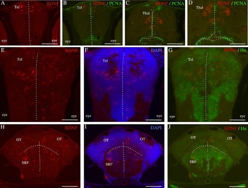
Immunohistochemical characterization of bdnf-expressing cells in the brain of 7 days old zebrafish larvae. Double staining for bdnf mRNA (red) and PCNA protein (green) on cross-sections through the telencephalon (A-B) and the thalamus (C-D). Double staining for bdnf mRNA (red) and the neuronal marker Hu (green) in the thalamus (E,F and G), the optic tectum (H, I and J) and at the level of the superior reticular formation (H, I and J). In F and I cells nuclei are counterstained with DAPI. The dotted lines indicate the ventricles. OT: optic tectum; SRF: superior reticular formation; Tel: telencephalon; Thal: thalamus. Scale bar: 150 µm in H, I and J. 120 µm in A, B, C. 60 µm in D, E, F and G. |
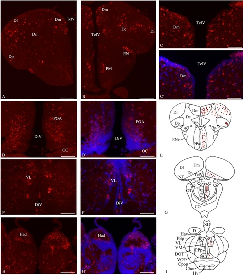
Bdnf mRNA distribution in cross-sections through the adult zebrafish forebrain. Telencephalon (A, B, C, C′ and E), preoptic area (B, D, D′ and E, G), thalamus (F, F′ and I) and dorsal habenula (H, H′). In C′, D′, F′, H′ cell nuclei are labeled in blue with DAPI. E, G and I are representative sections taken from the zebrafish atlas (Wullimann et al., 1996). Bdnf-expressing cells are represented by red dots. Dc: central zone of the dorsal telencephalon; DiV: diencephalic ventricle; Dl: lateral zone of the dorsal telencephalon; Dm: medial zone of the dorsal telencephalon; Dp: posterior zone of the dorsal telencephalon; EN: endopedoncular nucleus; Had: dorsal habenular nucleus; PM: magoncellular preoptic nucleus; OC: optic chiasma; POA: preoptic area; TelV: telencephalic ventricle; VL: ventrolateral thalamic nuclei. Scale bar: 120 µm except in C and C′: 60 µm. |

Bdnf mRNA distribution in cross-sections through the adult zebrafish mid- and hindbrain. In the ventral hypothalamus (A, A′, F and G), the dorsal thalamic region (B, B′), the optic tectum (C, C′ and G), the posterior tuberal nucleus (D, D′ and G) and medulla oblongata (E, E′ and H). In A′, B′, C′, D′ and E′, cell nuclei are labeled in blue with DAPI. F, G and H are representative sections taken from the zebrafish atlas (Wullimann et al., 1996). Bdnf-expressing cells are represented by red dots. DiV: diencephalic ventricle, DP: dorsal thalamic nucleus; Hd: dorsal zone of the periventricular hypothalamus; Hv: ventral zone of the periventricular hypothalamus; IRF: inferior reticular formation; OT: optic tectum; PGZ: periventricular gray zone of the optic tectum. PTN: posterior tuberal nucleus. Scale bar: 120 µm. |

Immunohistochemical characterization of bdnf-expressing cells in adult zebrafish brain. Double staining for bdnf mRNA (red) and PCNA protein (green) on cross-sections through the telencephalon (A to A′′′), the thalamus (B and B′) and the ventral hypothalamus (B and B′′). A and B are representative sections taken from the zebrafish atlas (Wullimann et al., 1996). Bdnf-expressing cells are represented by red dots and PCNA-labeled cells are green dots. Dl: lateral zone of the dorsal telencephalon; Dm: medial zone of the dorsal telencephalon; Hyp: hypothalamus; TelV: telencephalic ventricle; Thal: thalamus; Vv: ventral zone of the ventral telencephalon. Scale bar = 200 µm in A′; 100 µm in A′′; 50 µm in A′′′. |
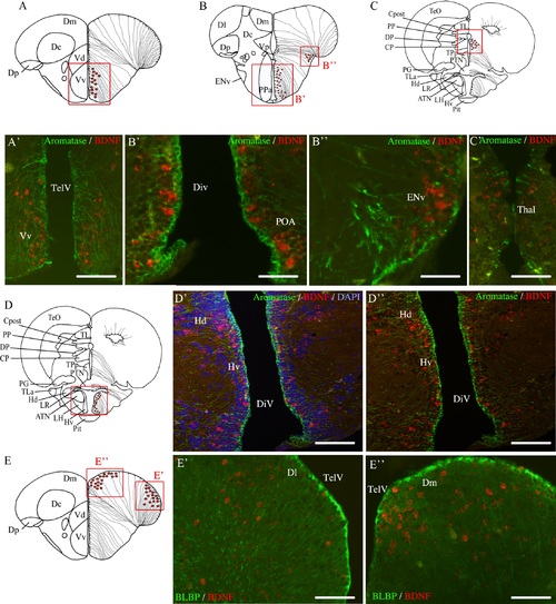
Immunohistochemical characterization of bdnf-expressing cells in adult zebrafish brain. A, B, C, D and E are representative sections taken from the zebrafish atlas (Wullimann et al., 1996). Bdnf-expressing cells are represented by red dots and Aromatase B (A-D) or BLBP-labeled cells (E) are represented by black dots with thin lines indicating radial glia cytoplasmic processes. Double staining for bdnf mRNA (red) and Aromatase B protein (green) on cross-sections through the telencephalon (A-A′), the preoptic area (B-B′), the entopedoncular nucleus (B-B′′), the thalamus (C-C′) and the ventral hypothalamus (D-D′′). Double staining for bdnf mRNA (red) and BLBP protein (green) on cross-sections through the telencephalon (E-E′′). DiV: diencephalic ventricle; Dl: lateral zone of the dorsal telencephalon; Dm: medial zone of the dorsal telencephalon; ENv: endopedoncular nucleus; Hd: dorsal zone of the periventricular hypothalamus; Hv: ventral zone of the periventricular hypothalamus; POA: preoptic area; TelV: telencephalic ventricle; Vv: ventral zone of the ventral telencephalon. D′ and D′′ are obtained with an Apotome-equipped Zeiss. Scale bar = 60 µm in A′, C′, D′, D′′, E′ and E′′; 30 µm in B′ and B′′. |
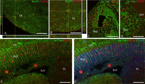
Transverse sections of adult zebrafish brain showing co-expression of bdnf mRNA with neuronal markers. Orthogonal projections of Z-stacks from telencephalon (11 sections of 0.5 µm) evidencing the expression of bdnf (red) in neurons identified by MAP2 protein (green) (A and B). Double staining for bdnf mRNA (red) and acetylated-tubuline (green) in the habenula (C), the thalamus (D) and the optic tectum (E-E′). In E′, cell nulei are labeled in blue with DAPI. Dl: lateral zone of the dorsal telencephalon; Had: dorsal habenular nucleus; OT: optic tectum; PGZ: periventricular gray zone of the optic tectum; TelV: telencephalic ventricle; Thal: thalamus; TL: torus longitudinalis. A, B, C and D were obtained with the confocal microscope. E and E′ were obtained with the Apotome. Scale bar: 50 µm in E and E′; 40 µm in A and C; 25 µm in B and D. |

The bdnf sense riboprobe did not generate any signal in the brain of 7 days old zebrafish. Telencephalon (A), Preoptic area (B), Optic Tectum (B and C). Hyp: hypothalamus; POA: preoptic area; OR: olfactory rosettes; OT: optic tectum. Teg: tegmentum; Tel: telencephalon. Scale bar: 120 µm in A and C. Scale bar: 60 µm in B. |
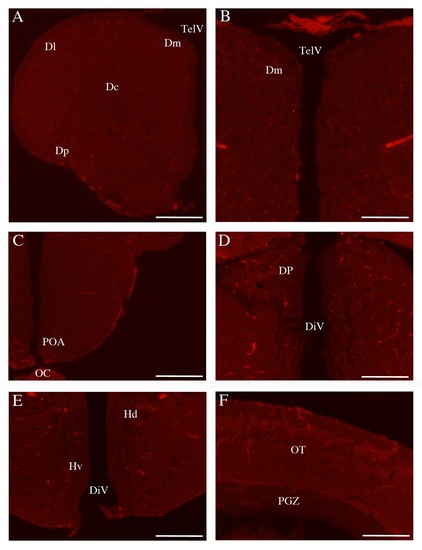
The bdnf sense riboprobe did not generate any signal in the brain of adult zebrafish. Telencephalon (A and B), Preoptic area (C), Thalamus (D), Hypothalamus (E) and Optic Tectum (F). Dc: central zone of the dorsal telencephalon; DiV: diencephalic ventricle; Dl: lateral zone of the dorsal telencephalon; Dm: medial zone of the dorsal telencephalon; Dp: posterior zone of the dorsal telencephalon; DP: dorsal thalamic nucleus; Hd: dorsal zone of the periventricular hypothalamus; Hv: ventral zone of the periventricular hypothalamus; OC: optic chiasma; OT: optic tectum; PGZ: periventricular gray zone of the optic tectum; POA: preoptic area. Scale bar: 120 µm in A, C. Scale bar: 60 µm in B, D, E, F. |