
- Title
-
Generation and characterization of FMR1 knockout zebrafish
- Authors
- den Broeder, M.J., van der Linde, H., Brouwer, J.R., Oostra, B.A., Willemsen, R., and Ketting, R.F.
- Source
- Full text @ PLoS One
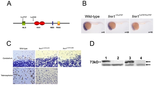
Fmr1 mutant alleles. A) Illustration of the fmr1 gene product. The different domains are indicated, along with the sites where isolated mutant alleles will affect the protein. B) Whole mount in situ hybridisation with an fmr1 specific probe. C) Immuno staining of wild type and mutant brain sections using Fmr specific antibodies. Some Purkinje cells in the mutant have been outlined. D) Brain lysates were analyzed by western blot, using an Fmr specific antibody. Lanes 1 and 3 contain wild type samples. Lane 2 contains hu2787/hu2787 lysate. Lane 4 contains hu2787/hu2898 lysate. The upper arrow points at Fmr. The lower arrow points at an a-specific band that serves as a loading control. EXPRESSION / LABELING:
|
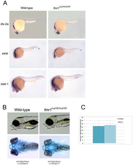
Phenotypic assays on wild-type and fmr1 mutant embryos. A) Wild type and mutant embryos were analyzed using whole mount in situ hybridisation using probes against dlx-2a, axial and islet-1. B) The width of Meckel′s cartilage was measured in wild type (n = 9) and MZ fmr1 mutant (n = 11) embryos. The angle of this structure with regard to the anterior-posterior axis was also measured in wild-type (n = 6) and fmr1 mutant (n = 9) embryos. Indicated errors represent SD. C) Neurite branching was measured on Rohon-Beard neurites using the monoclonal antibody zn-12. Plotted is the branching frequency per 1000 μm in both wild-type and MZ stop mutant embryos. In total n = 25 neurites (wild-type) and n = 28 neurites (MZfmr1) were traced in a total of 8 embryos of each genotype. Error bars represent SD. PHENOTYPE:
|

Unillustrated author statements PHENOTYPE:
|