
- Title
-
Expression of the eight AMPA receptor subunit genes in the developing central nervous system and sensory organs of zebrafish
- Authors
- Hoppmann, V., Wu, J.J., Søviknes, A.M., Helvik, J.V., and Becker, T.S.
- Source
- Full text @ Dev. Dyn.
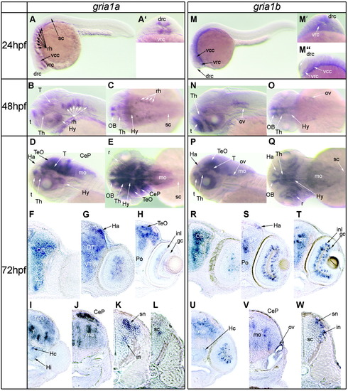
Expression of the paralogous AMPA receptor subunit genes gria1a and gria1b in zebrafish at 24, 48, and 72 hours postfertilization (hpf). A,A′: Gene expression of gria1a in a 24 hpf embryo (A: lateral view; A′: frontal view). B,C: The expression pattern of gria1a at 48 hpf (B: lateral view; C: dorsal view). D,E: Expression of gria1a at 72 hpf (D: lateral view; E: dorsal view). F-L: The 20-μm-thick, frontal vibratome sections through a gria1a hybridized larvae at 72 hpf. M-M″: Expression pattern of gria1b in a 24 hpf embryo (M: overview; M′: frontal view; M″: higher magnification of the forebrain in a lateral view). N,O: Expression of gria1b in the central nervous system of a 48 hpf zebrafish embryo (N: lateral view; O: dorsal view). P,Q: gria1b expression in a 72 hpf zebrafish larvae (P: lateral view; Q: dorsal view). R-W: Twenty-micrometer-thick Vibratome sections performed frontally through a gria1b hybridized zebrafish larvae at 72 hpf. CeP, cerebellar plate; drc, dorsal rostral cluster; DT, dorsal thalamus; gc, retinal ganglion cells; Ha, habenula; Hc, caudal hypothalamus; Hy, hypothalamus; in, area where inter neurons are located; inl, inner nuclear layer; mn, area where motor neurons are located; mo, medulla oblongata; OB, olfactory bulb; ov, otic vesicle; Po, preoptic region; r, retina; rh, rhombomeres; S, subpallium; sc, spinal cord; sn, sensory neurons; t, telencephalon; T, tegmentum; TeO, tectum opticum; Th, Thalamus; vcc, ventral caudal cluster; vrc, ventral rostral cluster; VT, ventral thalamus. EXPRESSION / LABELING:
|
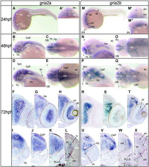
Expression of the paralogous AMPA receptor subunit genes gria2a and gria2b in zebrafish at 24, 48, and 72 hours postfertilization (hpf). A,A′: Expression of gria2a at 24 hpf (A: lateral view; A′: frontal view). B,C: The expression pattern of gria2a in the central nervous system (CNS) at 48 hpf (C: lateral view; D: dorsal view). D,E: Expression of gria2a at 72 hpf (D: lateral view; E: dorsal view). F-L: The 20-μm-thick frontal vibratome sections through a gria2a hybridized zebrafish larvae at 72 hpf. M-M′: Gene expression pattern of gria2b at 24 hpf (M: lateral view; M′: frontal view; M″: higher magnification of the forebrain in the lateral view). N,O: Expression of gria2b in the CNS of a zebrafish embryo at 48 hpf (N: lateral view; O: dorsal view). P,Q: gria2b expression in the CNS of a 72 hpf zebrafish larvae (P: lateral view; Q: dorsal view). R-W: The 20-μm-thick vibratome sections cut frontally through a gria2b hybridized 72 hpf zebrafish larvae. PLLG, posterior lateral line ganglion. Other abbreviations as in Figure 1. EXPRESSION / LABELING:
|

Expression of the paralogous AMPA receptor subunit genes gria3a and gria3b in zebrafish at 24, 48, and 72 hours postfertilization (hpf). A: gria3a expression at 24 hpf, lateral view. B,C: The expression pattern of gria3a at 48 hpf (B: lateral view; C: dorsal view). D,E: Expression of gria3a in the CNS in a 72 hpf zebrafish larvae (D: lateral view; E: dorsal view). F-L: The 20-μm-thick frontal Vibratome sections through a gria3a hybridized zebrafish larvae at 72 hpf. M: Gene expression of gria3b at 24 hpf, lateral view. N,O: Expression of gria3b in the central nervous system in a 48 hpf zebrafish embryo (N: lateral view; O: dorsal view). P,Q: gria3b expression in a 72 hpf zebrafish embryo (P: lateral view; Q: dorsal view). R-W: Frontal, 20-μm-thick Vibratome sections through a gria3b hybridized zebrafish larvae at 72 hpf. ALLG, anterior lateral line ganglion; hc, horizontal cells; OG, octaval ganglion; TG, trigeminal ganglion. Other abbreviations as in Figures 1 and 2. EXPRESSION / LABELING:
|
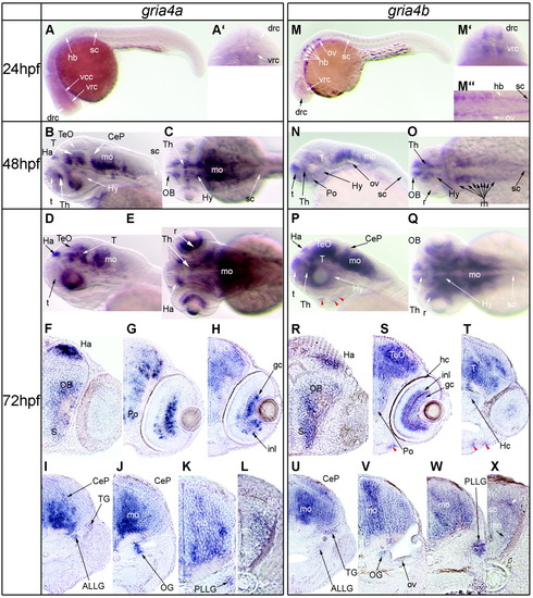
Expression of the paralogous AMPA receptor subunit genes gria4a and gria4b in zebrafish at 24, 48, and 72 hours postfertilization (hpf). A,A′: gria4a expression at 24 hpf (A: lateral view; A′: frontal view). B,C: The expression pattern of gria4a at 48 hpf (B: lateral view; C: dorsal view). D,E: Expression of gria4a in the central nervous system (CNS) of a 72 hpf zebrafish larvae (D: lateral view; E: dorsal view). F-L: The 20-μm-thick Vibratome sections cut frontally through a gria4a hybridized zebrafish larvae at 72 hpf. M-M″: gria4b gene expression pattern in a 24 hpf embryo (M: lateral view; M′: frontal view; M″: dorsal view with focus on hindbrain and spinal cord). N,O: Expression of gria4b in the CNS of 48 hpf zebrafish embryos (N: lateral view; O: dorsal view). P,Q: gria4b expression in a 72 hpf zebrafish larvae (P: lateral view; Q: dorsal view). R-W: Frontal, 20-μm-thick Vibratome sections performed through a gria4b hybridized zebrafish larvae at 72 hpf. For abbreviations, see Figures 1-3. EXPRESSION / LABELING:
|