
Fig. 3
- ID
- ZDB-IMAGE-240903-65
- Genes
- Publication
- Harboe et al., 2024 - The metalloproteinase PAPP-A is required for IGF-dependent chondrocyte differentiation and organization
- All Figures
- Figures for Harboe et al., 2024
|
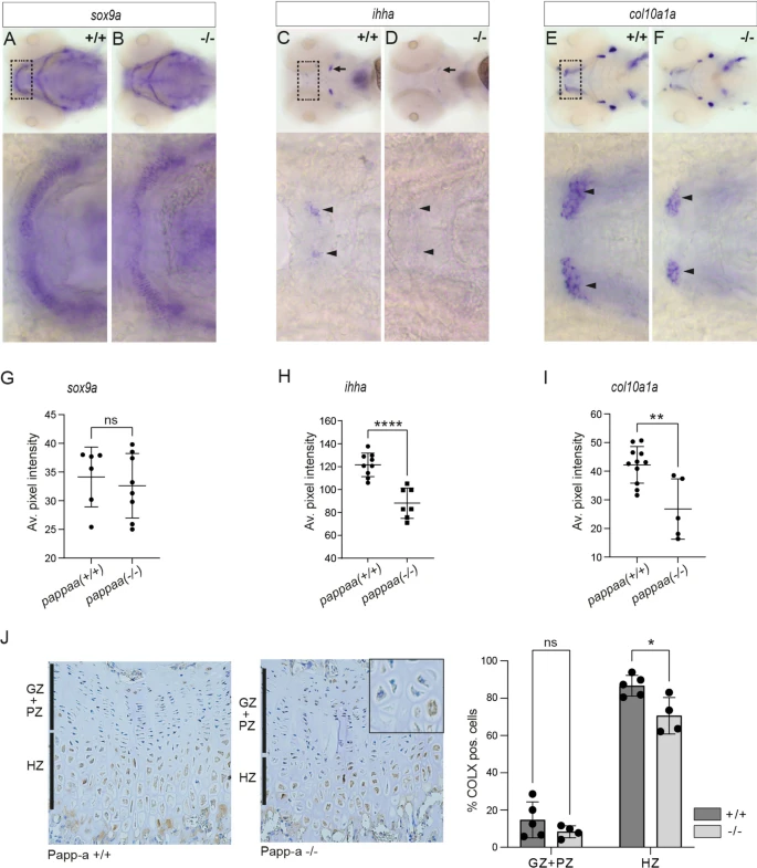
Fig. 3 Altered expression of chondrocyte differentiation markers sox9a, col10a1a and ihha in pappaa knockout larvae. At 4 dpf, the expression of sox9a, ihha, and col10a1 was assessed in wild-type and pappaa knockout larvae by whole mount in situ hybridization. (A,B) Expression of sox9a, a transcription factor expressed during early chondrogenesis essential for initiating and maintaining chondrogenesis26. (C,D) Expression of ihha, a paracrine factor synthesized by prehypertrophic chondrocytes27. Staining of ihha in the ceratohyal (arrows) or Meckel’s (arrowheads) cartilage is indicated. (E,F) Expression of col10a1a, an ECM component synthesized by hypertrophic chondrocytes27. In zebrafish, perichondral expression has also been reported around Meckel’s cartilage (arrowheads)28. (G–I) Average intensity of the staining in Meckel’s/ceratohyal cartilage for sox9a, ihha, and col10a1a, respectively. (J) Immunostaining of growth plate sections from wild-type or PAPP-A knockout mice, as indicated (left panels) using an antibody specific for mouse COLX. Quantification of the number of COLX-positive chondrocytes is shown for the germinal and proliferative zones combined (GZ + PZ) and for the hypertrophic zone (HZ) (right panel). An example of COLX-negative and -positive hypertrophic chondrocytes in the growth plate of PAPP-A knockout mouse is shown (inset).