
Fig. 3.
- ID
- ZDB-IMAGE-240518-19
- Genes
- Antibodies
- Publication
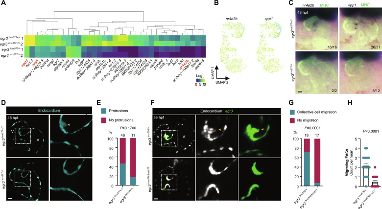
- da Silva et al., 2024 - egr3 is a mechanosensitive transcription factor gene required for cardiac valve morphogenesis
- All Figures
- Figures for da Silva et al., 2024
|
Fig. 3.
(