
Fig. 4
- ID
- ZDB-IMAGE-201124-1
- Genes
- Publication
- Frame et al., 2020 - Metabolic Regulation of Inflammasome Activity Controls Embryonic Hematopoietic Stem and Progenitor Cell Production
- All Figures
- Figures for Frame et al., 2020
|
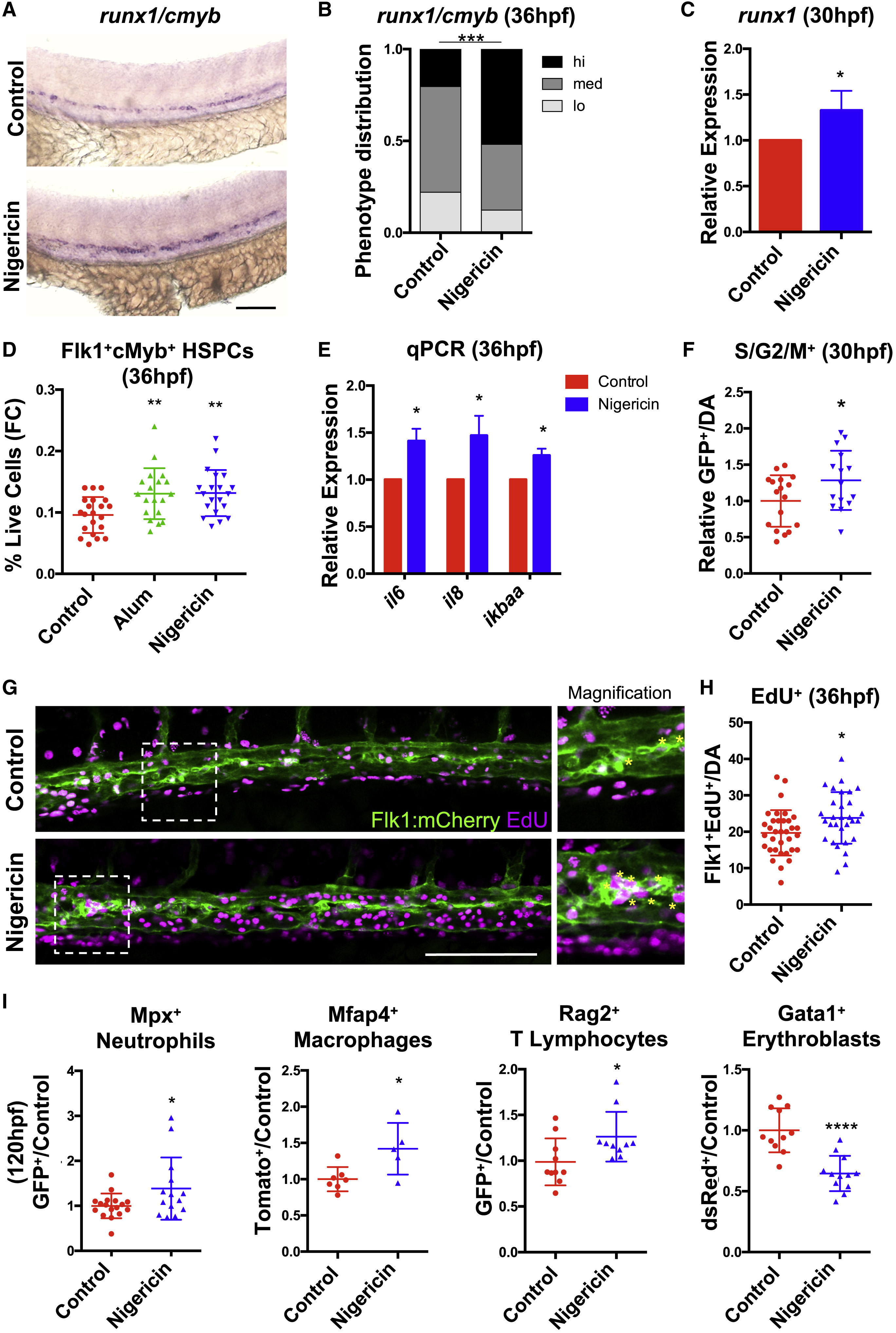
Fig. 4 HSPC Production Is Enhanced by Inflammasome Activation (A) Expression of runx1/cmyb in the dorsal aorta of embryos with or without inflammasome stimulation (12–36 hpf) assessed by WISH. (B) Phenotype distribution plot of runx1/cmyb expression scored in embryos from (A) (n ≥ 59/condition). (C) runx1 qPCR in embryos with or without inflammasome stimulation (n = 4, mean ±SEM). (D) Nigericin or Alum (12–36hpf) increased HSPCs by flow cytometry (FC). Significance was determined by ANOVA with Dunnett’s post-hoc test. Error bars indicate SD. (E) qPCR of IL1β target genes in embryos with or without inflammasome stimulation (n = 6, mean ± SEM). (F) Relative numbers of S/G2/M+ cells in Tg(EF1a:mAG-zGem(1/100)) aortas with nigericin stimulation (12–30 hpf) (DA, dorsal aorta; fold change calculated within each clutch). (G) Imaging of Flk1+EdU+ cells in the DA region in 36 hpf Tg(flk1:mCherry) embryos labeled with EdU antibody. Asterisks in inset signify positive cells in the aortic floor. (H) Quantification of Flk1+EdU+ cells in the aortic floor of embryos from (G). Error bars indicate SD. (I) The proportion of Mpx+ neutrophils, Mfap4+ macrophages, Rag2+ lymphocytes, and Gata1+ erythroblasts in transgenic embryos was assessed with prolonged Nigericin stimulation (24–120 hpf) by flow cytometry. Error bars indicate SD. ∗p < 0.05, ∗∗p < 0.01, ∗∗∗∗p < 0.0001. Scale bars, 100 μm. See also Figure S4.
Reprinted from Developmental Cell, 55(2), Frame, J.M., Kubaczka, C., Long, T.L., Esain, V., Soto, R.A., Hachimi, M., Jing, R., Shwartz, A., Goessling, W., Daley, G.Q., North, T.E., Metabolic Regulation of Inflammasome Activity Controls Embryonic Hematopoietic Stem and Progenitor Cell Production, 133-149.e6, Copyright (2020) with permission from Elsevier. Full text @ Dev. Cell