
Fig. 3
- ID
- ZDB-IMAGE-180705-9
- Genes
- Publication
- Liu et al., 2018 - Lipoprotein lipase regulates hematopoietic stem progenitor cell maintenance through DHA supply
- All Figures
- Figures for Liu et al., 2018
|
Fig. 3
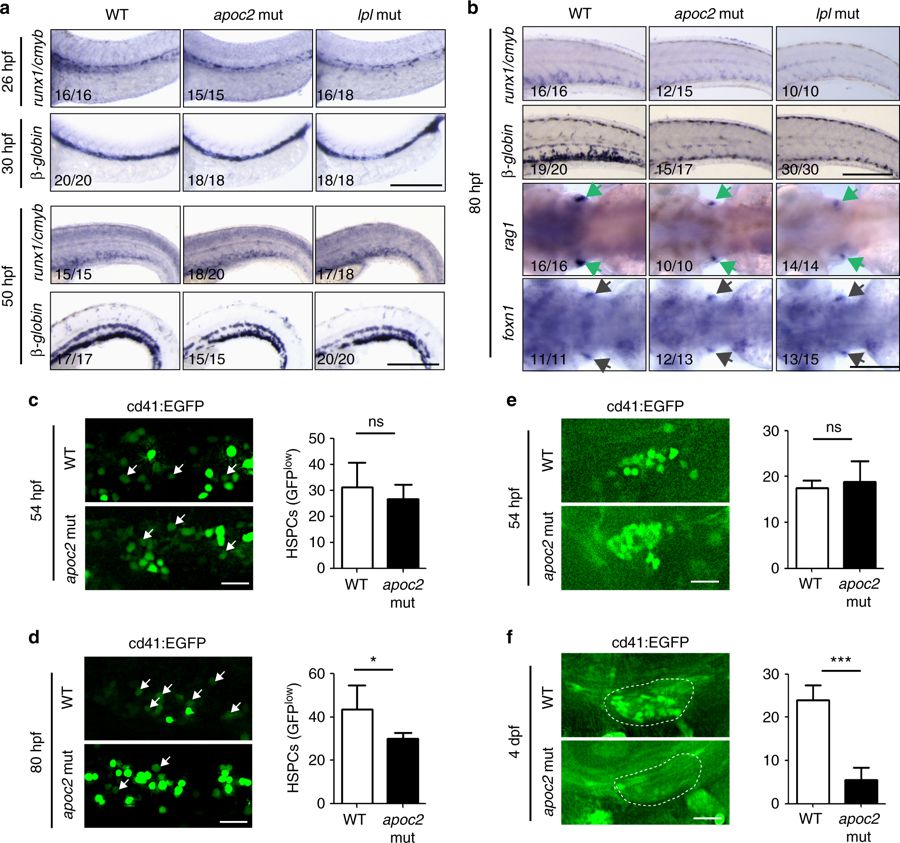
Hematopoietic defects in apoc2 and lpl mutants occur during HSPC expansion. a In situ hybridization with cmyb/runx1 and β-globin probes in WT, apoc2 and lpl mutants at 26 hpf, 30 hpf and 50 hpf. b In situ hybridization with cmyb/runx1, β-globin, rag1 (green arrows) and foxn1 (black arrows) probes in WT, apoc2 and lpl mutants at 80 hpf. foxn1 is a thymus development marker, used as a control. c, d Representative images and numbers of GFPlow cells (HSPCs, white arrows) in the CHT region of cd41:EGFP transgenic WT and apoc2 mutants at 54 and 80 hpf (n = 10 in WT and n = 8 in apoc2 mutant groups at 54 hpf; n = 6 in each group at 80 hpf). e, f Representative images and numbers of GFP-positive cells in the thymus region at 54 hpf (n = 9 in WT and n = 8 in apoc2 mutant groups) and at 4 dpf (n = 8 in each group). Scale bars, 200 μm in a and b; 50 μm in c–f. Mean ± SEM; *P < 0.05 and ***P < 0.001 (Student’s t test)