
Fig. 4
- ID
- ZDB-IMAGE-160915-40
- Genes
- Publication
- Mullins et al., 1996 - Genes establishing dorsoventral pattern formation in the zebrafish embryo: the ventral specifying genes
- All Figures
- Figures for Mullins et al., 1996
|
Fig. 4
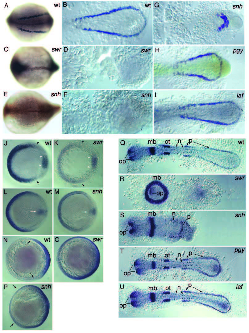
Altered expression of gata1, eve1, fkd3 and pax2 in whole-mount in situ hybridizations of the dorsalized mutants. Expression of gata1 in 8-somite stage wild-type embryos (A,B), swr (C,D), snh (E,F,G), pgy (H), and laf (I) mutant embryos. Double stainings with gata1 and anti-Ntl antibody are shown as whole mounts in A,C,E. Spreads are shown in (B,D,F-I). eve1, gsc double staining at shield stage in wild-type (J) and swr mutant (K) embryos (white arrowheads delineate gsc expression). eve1, gsc double stainings at 70% epiboly in wild-type (L), and snh mutant (M) embryos (white arrow marks gsc expression). fkd3 expression at 70% epiboly in wild-type (N), swr mutant (O), and snh mutant (P) embryos (black arrows indicate the lateral extent of staining). Spreads of pax2-stained 10-somite stage wild type (Q), swr (the most anterior position of the embryo lies in the middle of the ring; R), snh (S), pgy (T), and laf (U) mutant embryos. op, optic vesicle and stalk; mb, midbrain; ot, otic vesicle; n, neuronal and p, pronephric precursor expression domains.