
Fig. 5
- ID
- ZDB-IMAGE-160818-47
- Genes
- Publication
- Muthu et al., 2016 - Rx3 and Shh direct anisotropic growth and specification in the zebrafish tuberal/anterior hypothalamus
- All Figures
- Figures for Muthu et al., 2016
|
Fig. 5
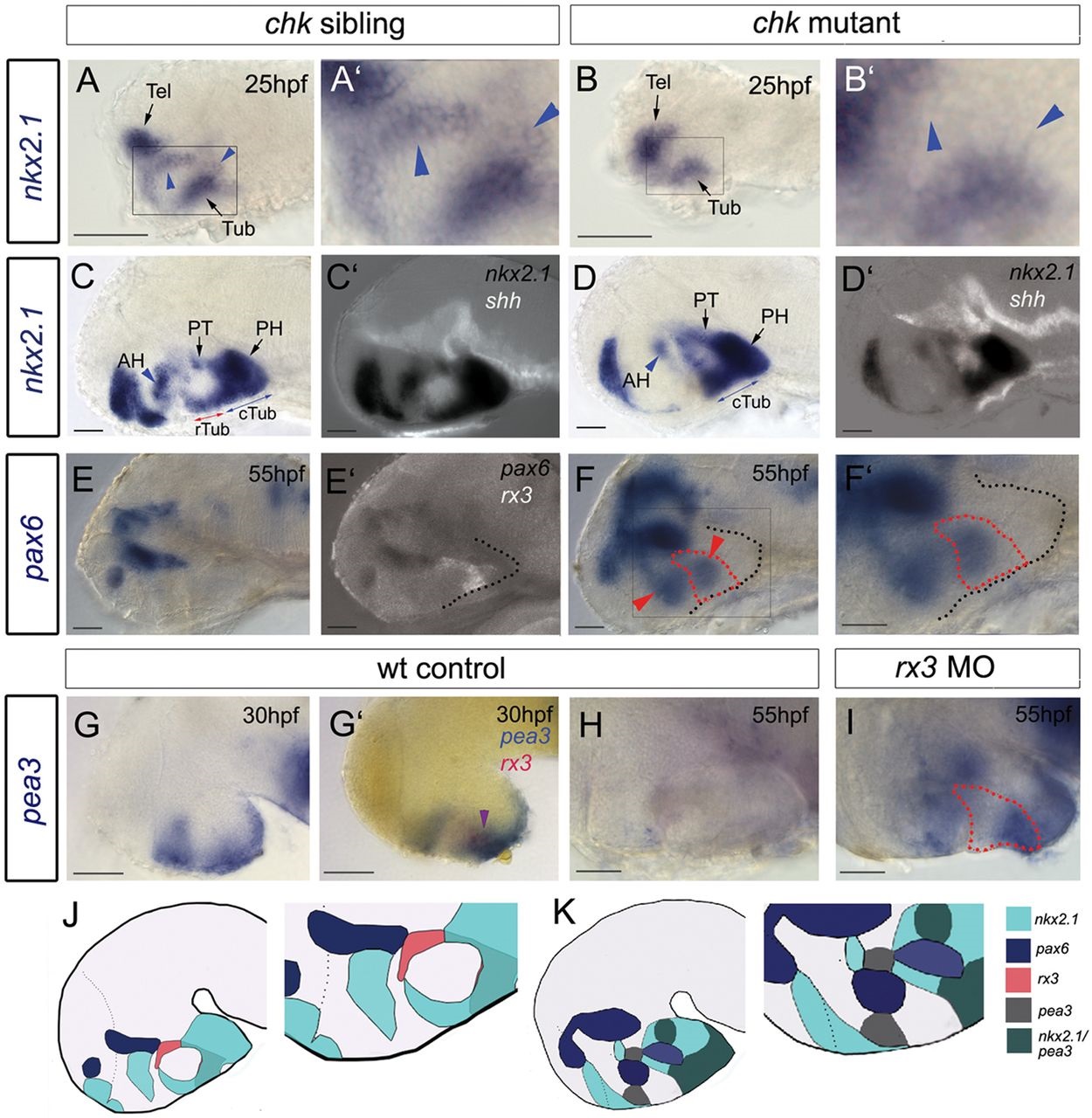
Rx3 suppresses dorsal and ventro-tuberal progenitors. (A-I) Side views of control embryos or embryos in which rx3 is absent. A′,B′,F′ show high-power views of boxed regions in A,B,F. Blue arrowheads and red arrows in A-D point to nkx2.1+ cells, which are absent in chk mutants. Blue arrows in C,D point to nkx2.1+ ventral-tuberal domain. Red arrowheads in F point to ectopic pax6+ cells. Black dotted lines indicate outline of ventral hypothalamus. Red dotted lines as in Fig. 2. Purple arrowhead in G′ points to rx3+pea3+ cells. H,I show views of isolated neuroectoderm. (J,K) Schematics of expression patterns in chk sibling (J) or mutant (K) 55hpf embryos. White and red dotted lines as in Fig. 2 AH, anterior hypothalamus; PH, posterior hypothalamus; PT, posterior tuberculum; Tel, telencephalon; (c)(r)Tub, (caudal) (rostral) tuberal hypothalamus. Scale bars: 50µm.