
Fig. 3
- ID
- ZDB-IMAGE-160818-44
- Genes
- Publication
- Muthu et al., 2016 - Rx3 and Shh direct anisotropic growth and specification in the zebrafish tuberal/anterior hypothalamus
- All Figures
- Figures for Muthu et al., 2016
|
Fig. 3
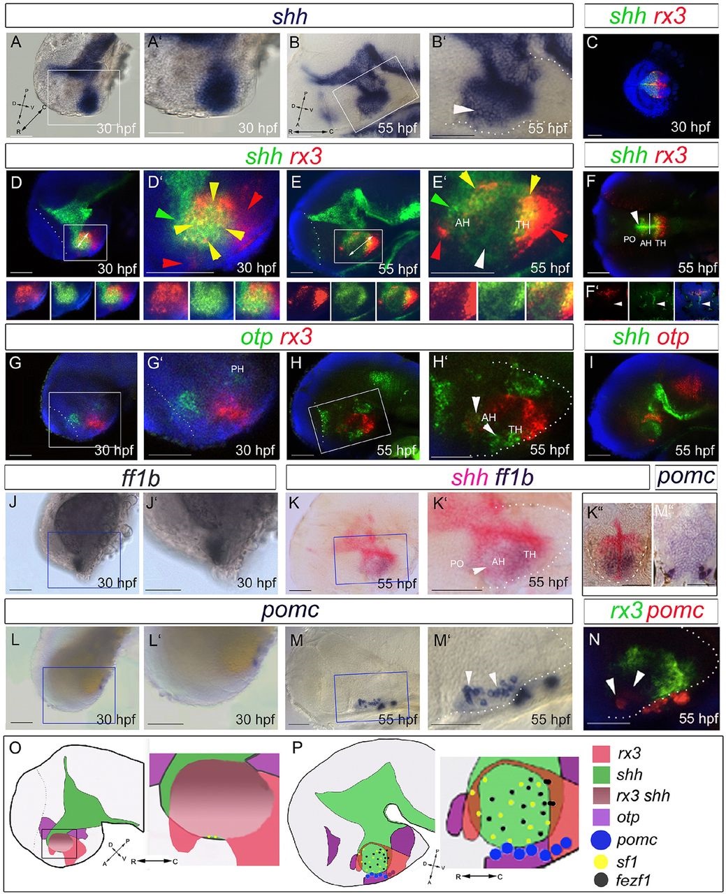
Differentiation in the 30-55hpf anterior/tuberal hypothalamus. (A-N) Side views (A,B,D,E,G-J,L-N), ventral views (C,F), sagittal (K) or transverse (K′′,M′′) sections of 30hpf and 55hpf embryos. A′,M′ show high-power views of boxed regions. In B′,E′,F, white arrowheads point to shh(weak+) AR cells; in H′, to otpb+ cells in the tuberal/anterior hypothalamus; in M′,N, to hypothalamic pomc+ cells. In D′,E′, arrowheads point to rx3+shh+ cells (yellow), rx3+ cells (red) or shh+ cells (green). (O,P) Schematics depicting expression domains at 30hpf (O) or 55hpf (P). AH, anterior hypothalamus; PH, posterior hypothalamus; PO, preoptic hypothalamus; TH; tuberal hypothalamus. Scale bars: 50µm.