
Fig. 9
|
Fig. 9
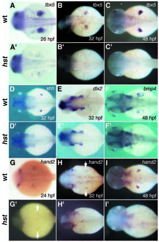
Fin bud development in hst mutant embryos. (A-I) Wild-type embryos, (A′-I′) hst mutant embryos. (A-C′) tbx5 expression in the developing pectoral fin field. At 26 hpf, (A) wild-type embryos restrict tbx5 expression to a circular patch, the presumptive fin field, in the dorsal LPM, but (A′) hst mutant embryos display a more dispersed, less abundant expression. (B,B′) At 32 hpf and (C,C′) at 48 hpf, (B,C) wild-type embryos continue tbx5 expression in the developing bud mesenchyme, but (B′,C′) in hst mutant embryos, tbx5 expression is greatly decreased (B′) at 32 hpf and (C′) undetectable in the LPM thereafter. (D-F′) Expression of molecular markers of early limb bud induction. (D,D′) At 32 hpf, wild-type embryos express shh expression in the pectoral fin mesenchyme, but hst mutant embryos do not. (E,E′) Likewise, at 32 hpf dlx2 is expressed in the apical fold of wild-type but not hst mutant embryos. (F,F′) At 48 hpf, bmp4 is present in the apical fold of wild-type but not hst mutant embryos. (G-I′) hand2 expression in the developing fin field. (G,G′) At 24 hpf, wild-type embryos express hand2 broadly in the region of the LPM encompassing the fin field, but hst mutant embryos express hand2 faintly in only a few LPM cells. (H,H′) At 32 and (I,I′) at 48 hfp, wild-type embryos (H,I) express hand2 in the fin bud mesenchyme, but hst mutant embryos (H′,I′) have no detectable hand2 expression in the LPM. Arrows in H and G′ indicate cells weakly expressing hand2.