
Fig. 5
- ID
- ZDB-IMAGE-151117-12
- Genes
- Publication
- Wu et al., 2015 - Control of Wnt5b secretion by wntless modulates chondrogenic cell proliferation through fine-tuning fgf3 expression
- All Figures
- Figures for Wu et al., 2015
|
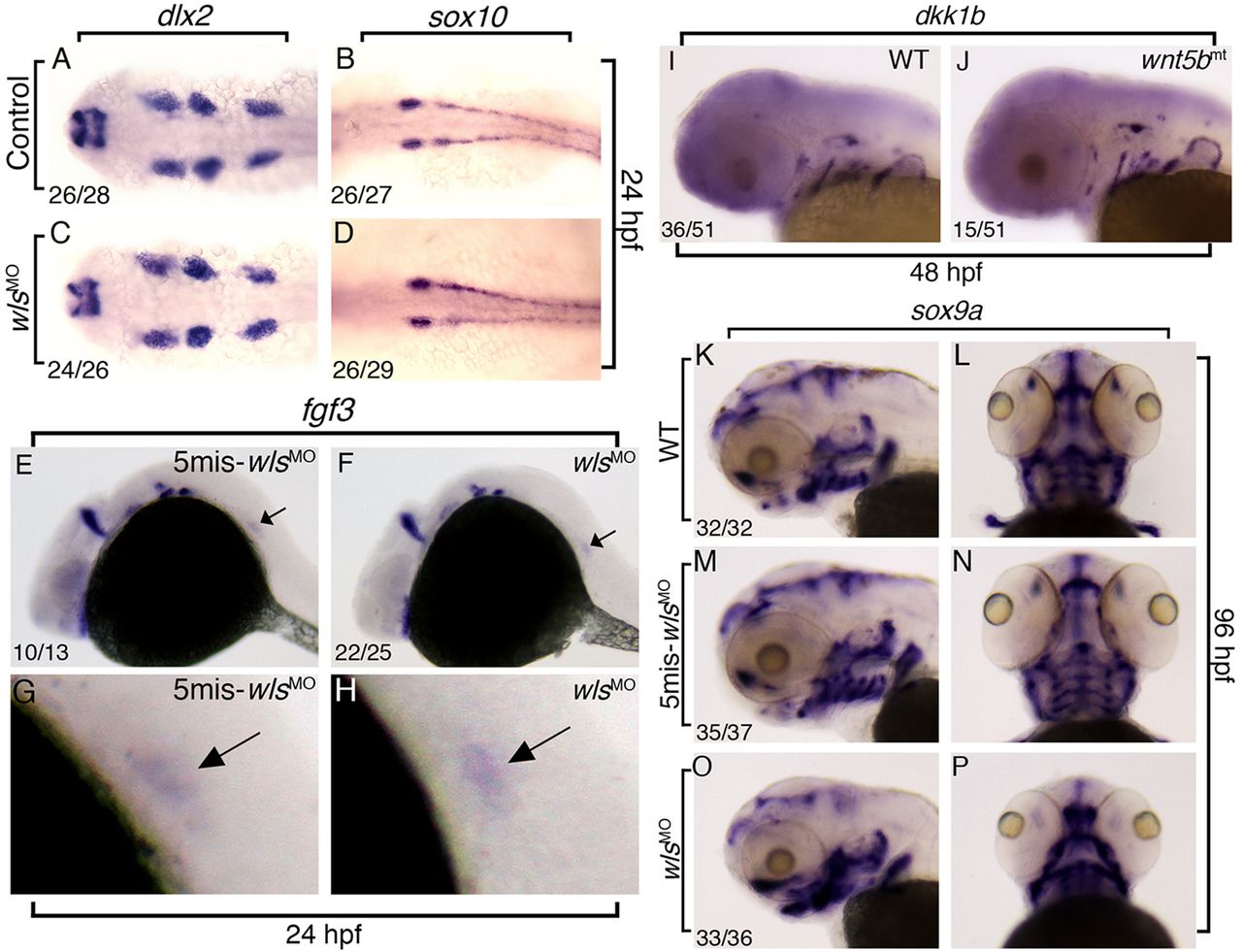
Fig. 5 Migration and specification of neural crest cells are normal in wls morphants. (A–D) Dorsal views of 24 hpf control (5mis-wlsMO) (A,B) or wls-MO-injected (wlsMO) (C,D) embryos labeled with an antisense probe for dlx2 (A,C) or sox10 (B,D). (E–H) Lateral views of 24 hpf 5mis-wlsMO (in E) or wlsMO (in F) embryos labeled with the antisense fgf3 probes. Enlarged trunk regions from E and F are shown in G and H. Arrows indicate the migrating lateral line promodia. (I,J) Lateral views of 48 hpf WT (in I) or wnt5bmt (in J) embryos labeled with antisense dkk1b probes. (K–P) Images showing the sox9a expression in WT (K,L), wls 5mis-MO-injected (wlscMO) (M,N) and wlsMO (O,P) larvae at 96 hpf. Lateral views are shown in K,M,O; ventral views are shown in L,N,P. Numbers in the bottom left corners indicate the number of the embryos showing the indicated phenotype and the total number examined (denominator).