
Fig. 5
- ID
- ZDB-IMAGE-140317-18
- Genes
- Publication
- Artinger et al., 1999 - Zebrafish narrowminded suggests a genetic link between formation of neural crest and primary sensory neurons
- All Figures
- Figures for Artinger et al., 1999
|
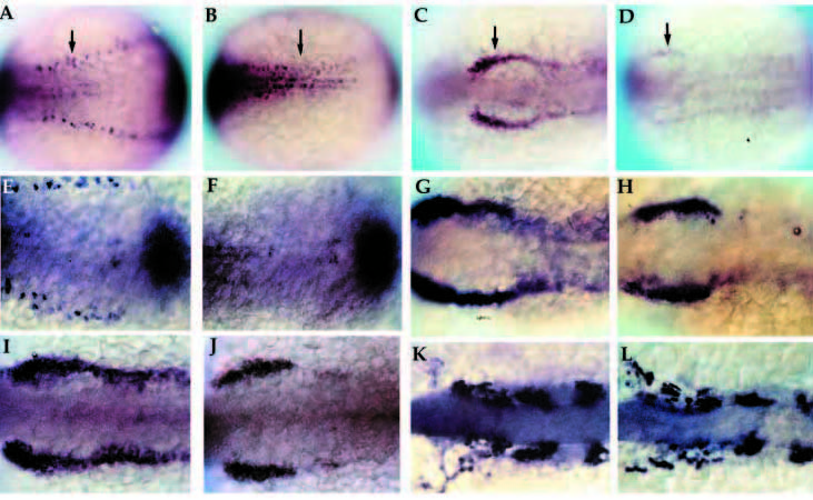
Fig. 5 Early development of primary neurons and neural crest in nrd. Wholemount in situ hybridization of 2-3 somite stage zebrafish embryos with HuC (A,B; 20x), snail2 at the 2 somite stage (C,D; 20x), BMP-4 (E,F; 32x), snail2 at the 4-5 somite stage (G,H; 32x), fkd6 at the 2-3 somite stage (I,J; 32x), and dlx2 at the 10 somite stage (K,L; 32x). All images show dorsal views of embryos orientated anterior to the left. Wildtype (A,C,E,G,I,K) and nrd mutant (B,D,F,H,J,L) embryos. nrd embryos shows a lack of HuC expression in the lateral most stripe of primary neurons, corresponding to the lateral edge of the neural plate (arrows in A,B). snail2 expression indicates a severe reduction in the neural crest population in the mutant embryos (arrows in C,D). This is also consistant with effects on the neural crest expression domain of fkd6, which appears reduced in nrd although less severe than snail2 (I,J). BMP-4 expression appears normal in nrd (compare E and F; double stain with HuC to identify mutant embryos). Later in development, at the 4-5 somite stage, snail2 expression appears to be upregulated again (compare nrd, in H, to wild type, in G) and is almost indistinguishable from wild type at the 10 somite stage, shown here with dlx2 expression (K,L).