
|
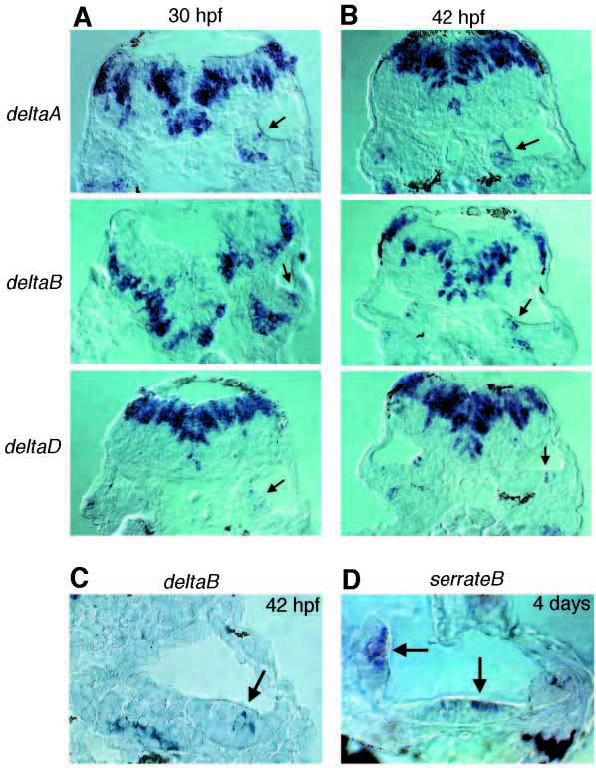
Fig. 3 (A,B) Expression of deltaA, deltaB and deltaD in wild-type ears at 30 and 42 hpf, shown by in situ hybridisation on cryosections cut transverse to the main body axis. Arrows indicate scattered cells expressing delta genes in the thickened ventral wall of the otocyst. Beneath this epithelium, in the 30 hpf deltaA and deltaB sections, one can see delta gene expression in cells of the statoacoustic ganglion. (C) Expression of deltaB in a cell in the hair-cell layer of a sensory patch (the anteroventral macula) at 42 hpf; such deltaB-expressing hair cells are few and far between, because expression of the gene fades rapidly as a hair cell matures. (D) Persistent and extensive expression of serrateB, seen at 4 days in the hair-cell layer of the posteromedial and anteroventral maculae (arrows).