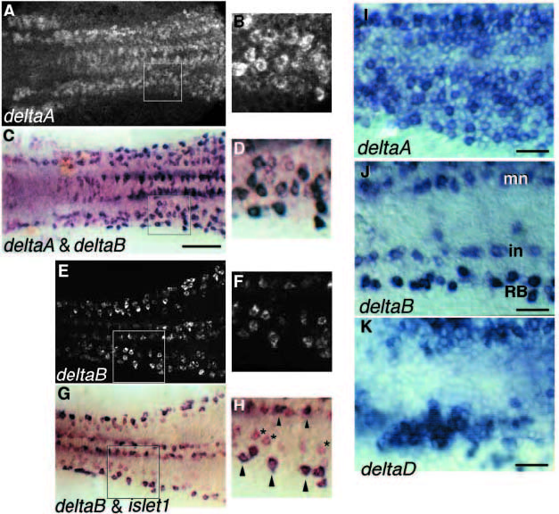
Fig. 5 (A-D) Coexpression of deltaA and deltaB in the same cells in the neural plate at the 5-somite stage. The boxed regions on the left are shown enlarged on the right (B,D). The cells expressing deltaB are generally the same that express deltaA most strongly. The embryo was first stained for deltaA expression, revealed by in situ hybridisation using fluorescent Fast Red detection and was viewed intact by epifluorescence, using the confocal microscope to construct an extended-focus image. The same specimen was then hybridised with a probe for deltaB, revealed in purple with NBT/BCIP, and was flatmounted and photographed with bright-field optics. Sequential imaging was used because the dark NBT/BCIP stain often obscures the Fast Red fluorescence. (E-H) Coexpression of deltaB and islet1 in the neural plate at 5 somites, shown by double in situ hybridisation. deltaB in red (fluorescence in upper panel (E,F), bright field in lower panel (G,H)), islet1 in blue-black (bright field, lower panel). Primary motor neurons and Rohon-Beard cells express both genes (arrowheads); cells expressing deltaB but not islet1 are probably primary interneurons (asterisks). (I-K) Details of the prospective anterior spinal region at the 5-somite stage, showing the diffuse but uneven expression of deltaA and deltaD in large patches of contiguous cells, and the more restricted expression of deltaB in scattered, isolated cells. In each case, the left side of the neural plate is shown, with midline at the top and anterior to the left of the picture. Scale bars: 100 μm (A-H), 50 μm (I-K).
Image
Figure Caption
Figure Data
Acknowledgments
This image is the copyrighted work of the attributed author or publisher, and
ZFIN has permission only to display this image to its users.
Additional permissions should be obtained from the applicable author or publisher of the image.
Full text @ Development

