
Fig. 7
- ID
- ZDB-IMAGE-140228-39
- Genes
- Publication
- Stainier et al., 1995 - cloche, an early acting zebrafish gene, is required by both the endothelial and hematopoietic lineages
- All Figures
- Figures for Stainier et al., 1995
|
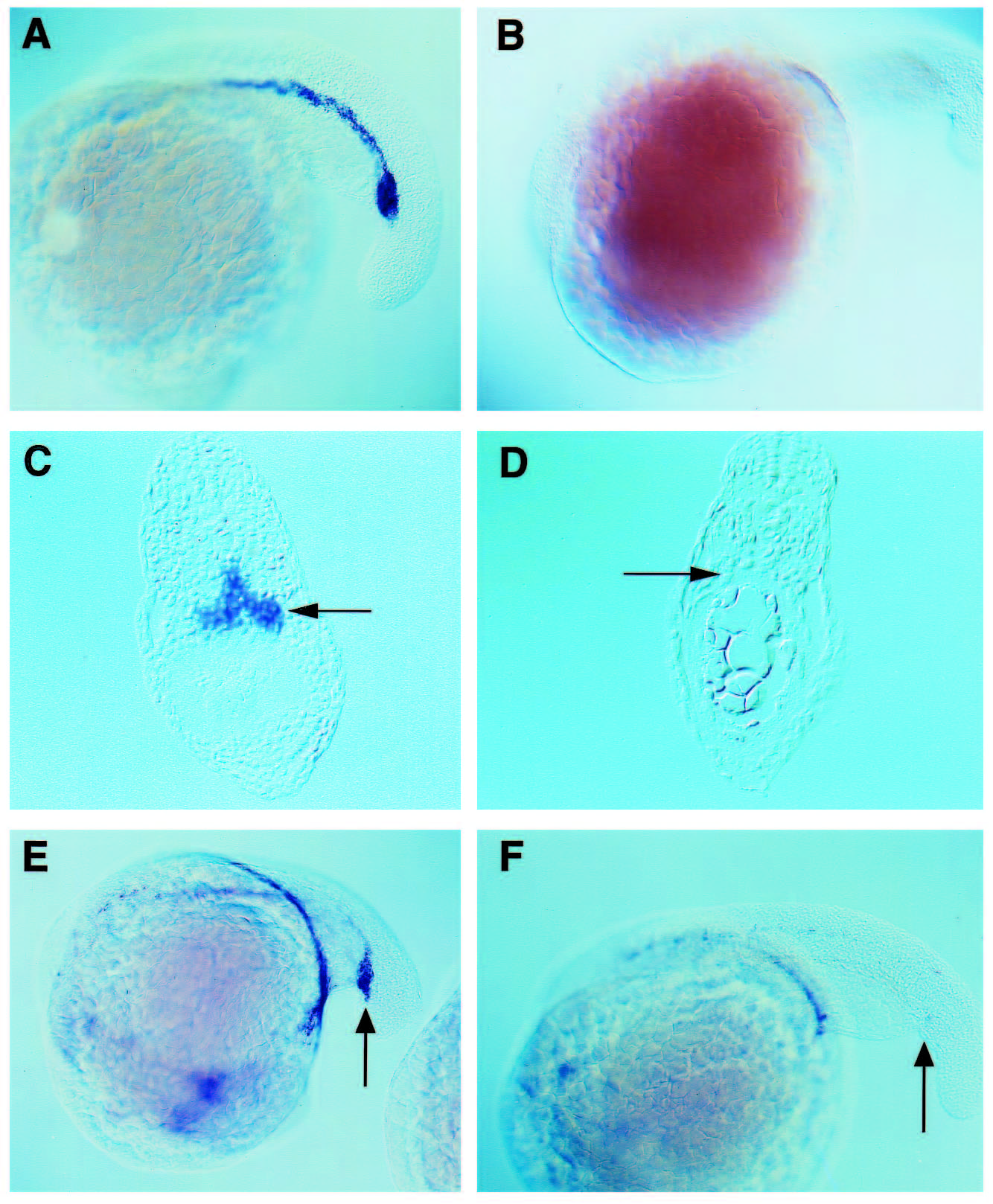
Fig. 7 clo mutant embryos do not express GATA-1 or GATA-2 in their hematopoietic tissues. In situ hybridization with a GATA-1 or GATA-2 antisense RNA probe was performed on wild-type embryos (A,C,E) and on clo mutants (B,D,F). (A-D) GATA-1 staining at the 22-somite stage. In wild-type embryos, GATA-1 expression is originally found in two longitudinal chords of cells that extend from the mid-trunk region to just beyond the yolk tube and pronephric ducts. These cell populations will fuse at the midline to form the ICM as seen in whole mounts (A) and transverse sections of the tail region (C). clo mutants fail to express GATA-1 (B,D). (E,F) GATA-2 staining of 20-somite stage embryos. GATA-2 is normally expressed from an early stage in all hematopoietic progenitors including the presumptive stem cells of the posterior ICM (E, arrow). In clo mutants, the hematopoietic tissues do not express GATA-2 (F). GATA-2 is also expressed in certain neural tissues (Detrich et al., 1995) and this expression is normal in mutant embryos (data not included).