
|
Fig. 1
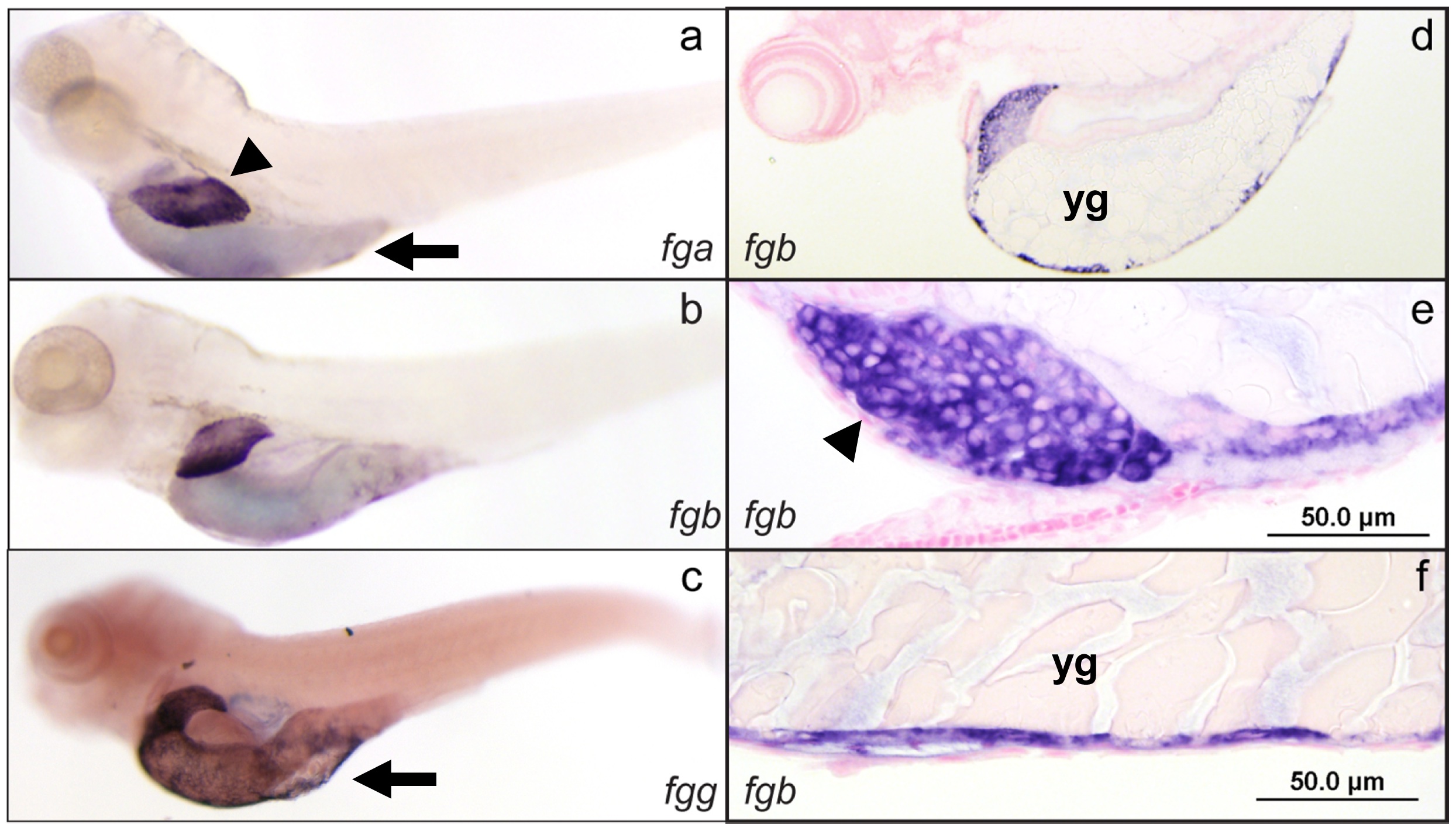
In situ hybridization of zebrafish fibrinogen genes demonstrates liver and yolk sac specific expression.
Whole mount in situ hybridization was performed using antisense mRNA probes complementary to the fga, fgb, and fgg mRNAs (a, b, and c respectively) at 5 dpf. The expression patterns were identical, with high levels in the liver and yolk sac. Individual experiments showed variability in yolk sac expression for all 3 genes, e.g. lower (a, b) and higher (c) levels of expression. Sections through fgb probed larvae confirmed that liver expression was in hepatocytes, while the yolk sac staining was in the outer cellular layer (d–f). Arrowheads indicate the liver, while arrows point to the yolk sac; yg, yolk granules. Sections of fga and fgg probed larvae demonstrated the same pattern, while in situ hybridization with probes generated from the sense strand did not show any signal (not shown).