
|
Fig. 1
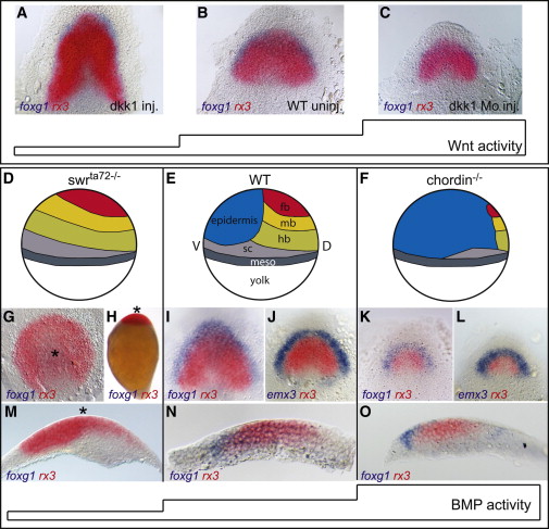
BMP, but Not Wnt, Subdivides the Anterior Forebrain Neural Fate into Eye and Telencephalon(A–C) Foxg1/rx3 in situ hybridization of dkk1 RNA (A) and dkk1 morpholino-injected embryos (C) compared to uninjected control (B) bud stage embryos (end of gastrulation), dorsal view, anterior to the top.(D–F) Schematic illustration of fate changes in response to BMP depletion in swrta72-/- (D) and elevated level of BMP signaling in chordin/ (F), compared to wild-type (E) bud stage embryos, lateral view, dorsal to the right. fb, forebrain; mb, midbrain; hb, hindbrain; sc, spinal chord; meso, mesoderm.(G–O) Foxg1, rx3 double in situ hybridization in swrta72-/- (G, H, and M), WT (I, J, and N), and chordin/ (K, L, and O). Asterisks in (G), (H), and (M) mark the center of the radialized axis. (G) Animal pole view, (H) lateral view, (I–L) dorsal view, anterior to the top, (M–O) sagittal section of the neural plate, anterior to the left.
Reprinted from Developmental Cell, 23(4), Bielen, H., and Houart, C., BMP Signaling Protects Telencephalic Fate by Repressing Eye Identity and Its Cxcr4-Dependent Morphogenesis, 812-822, Copyright (2012) with permission from Elsevier. Full text @ Dev. Cell