
Fig. S1
- ID
- ZDB-IMAGE-120810-47
- Genes
- Publication
- Hutchinson et al., 2012 - Tbl3 regulates cell cycle length during zebrafish development
- All Figures
- Figures for Hutchinson et al., 2012
|
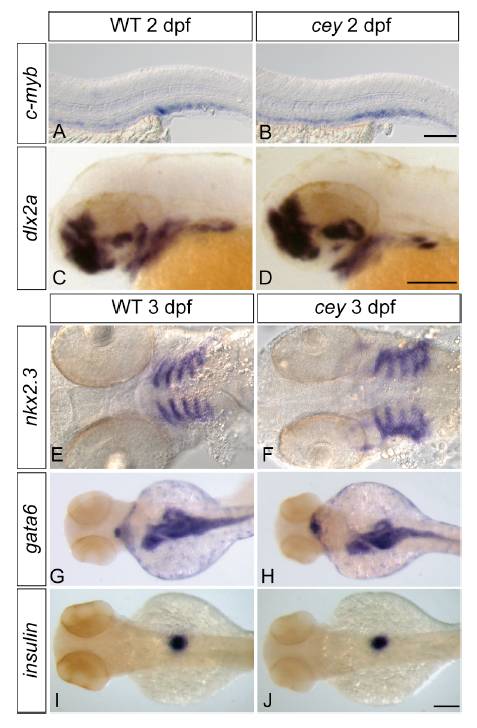
Fig. S1 Tissue specification and patterning is normal in cey mutants
Despite the severe reduction of HSPCs in cey mutants at 4 dpf, HSPCs labeled by c-myb in cey mutants at 2 dpf are comparable to wild-type (A, B). The cranial neural crest labeled by dlx2a gives rise to jaw cartilage and is normal at 2 dpf in cey mutants (C, D). At 3 dpf the endoderm that contributes to jaw formation is specified properly as indicated by nkx2.3 staining in cey mutants (E, F). Patterning and size of the intestinal and pancreatic anlage is also normal as shown by gata6 staining in cey mutants at 3 dpf (G, H). While differentiation of the exocrine pancreas is deficient, beta cells in the endocrine pancreas labeled by insulin are normal in cey mutants at 3 dpf (I, J). Scale bars are 125 μm.
Reprinted from Developmental Biology, 368(2), Hutchinson, S.A., Tooke-Locke, E., Wang, J., Tsai, S., Katz, T., and Trede, N.S., Tbl3 regulates cell cycle length during zebrafish development, 261-272, Copyright (2012) with permission from Elsevier. Full text @ Dev. Biol.