
Fig. 7
- ID
- ZDB-IMAGE-120216-90
- Genes
- Antibodies
- Publication
- Wu et al., 2011 - Ryanodine receptors, a family of intracellular calcium ion channels, are expressed throughout early vertebrate development
- All Figures
- Figures for Wu et al., 2011
|
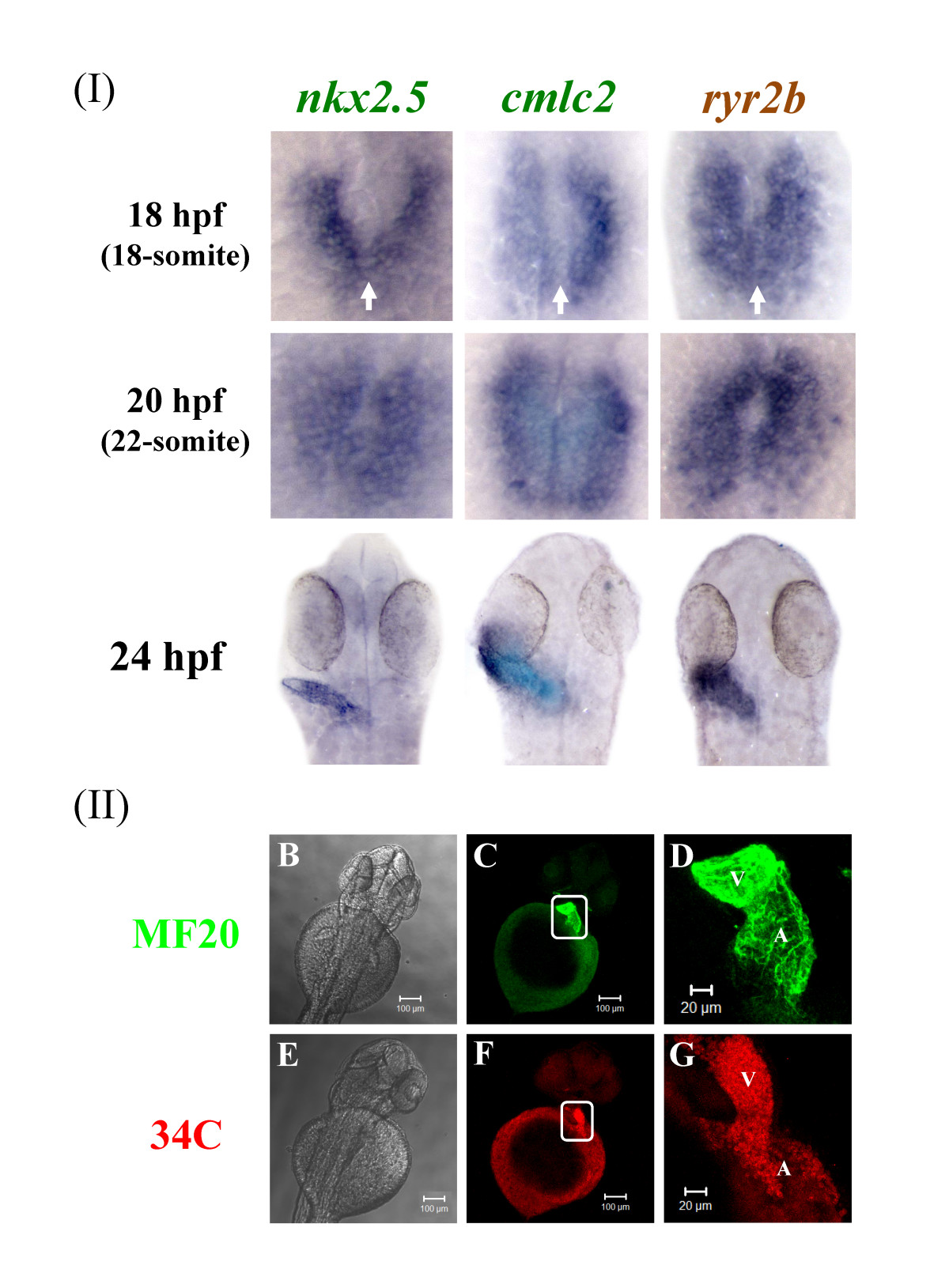
Fig. 7
Zebrafish ryr2b mRNA is expressed during early heart formation and the RyR protein throughout the heart at later stages. (a) Expression of nkx2.5, cmlc2, and ryr2b in 18 hpf (18-somite stage) was observed in the bilateral cardiac primordial cells. At this stage the bilateral cardiac primordial cells make contact and begin to fuse (arrows). The posterior portions fuse initially followed by anterior portions to create a central lumen and cardiac cone by 20 hpf (22-somite stage). A linear heart tube has formed by 24 hpf and ryr2b and cmlc2+ cells are expressed throughout the heart at this stage. 18- and 22-somite stage embryos, in which the tail was removed but the yolk sac left intact, were orientated so that anterior is to the top. Dorsal view of flat mounted embryos at 24 hpf. (b&e) Brightfield images of the anterior portion of the zebrafish embryo with the yolk sac still intact and dorsal side uppermost. Embryos were fixed and stained at 48 hpf to reveal either myosin (c-d) or RyRs (f-g), the images within the white boxes are shown in greater detail (d & g). By 48 hpf RyRs are expressed throughout the two chambers, atrium (A) and ventricle (V) of the heart. Images were taken using X10 (b,c & e,f) and X20 (d & g) objectives.