
Fig. 1
- ID
- ZDB-IMAGE-110915-14
- Genes
- Antibodies
- Publication
- Totong et al., 2011 - The novel transmembrane protein Tmem2 is essential for coordination of myocardial and endocardial morphogenesis
- All Figures
- Figures for Totong et al., 2011
|
Fig. 1
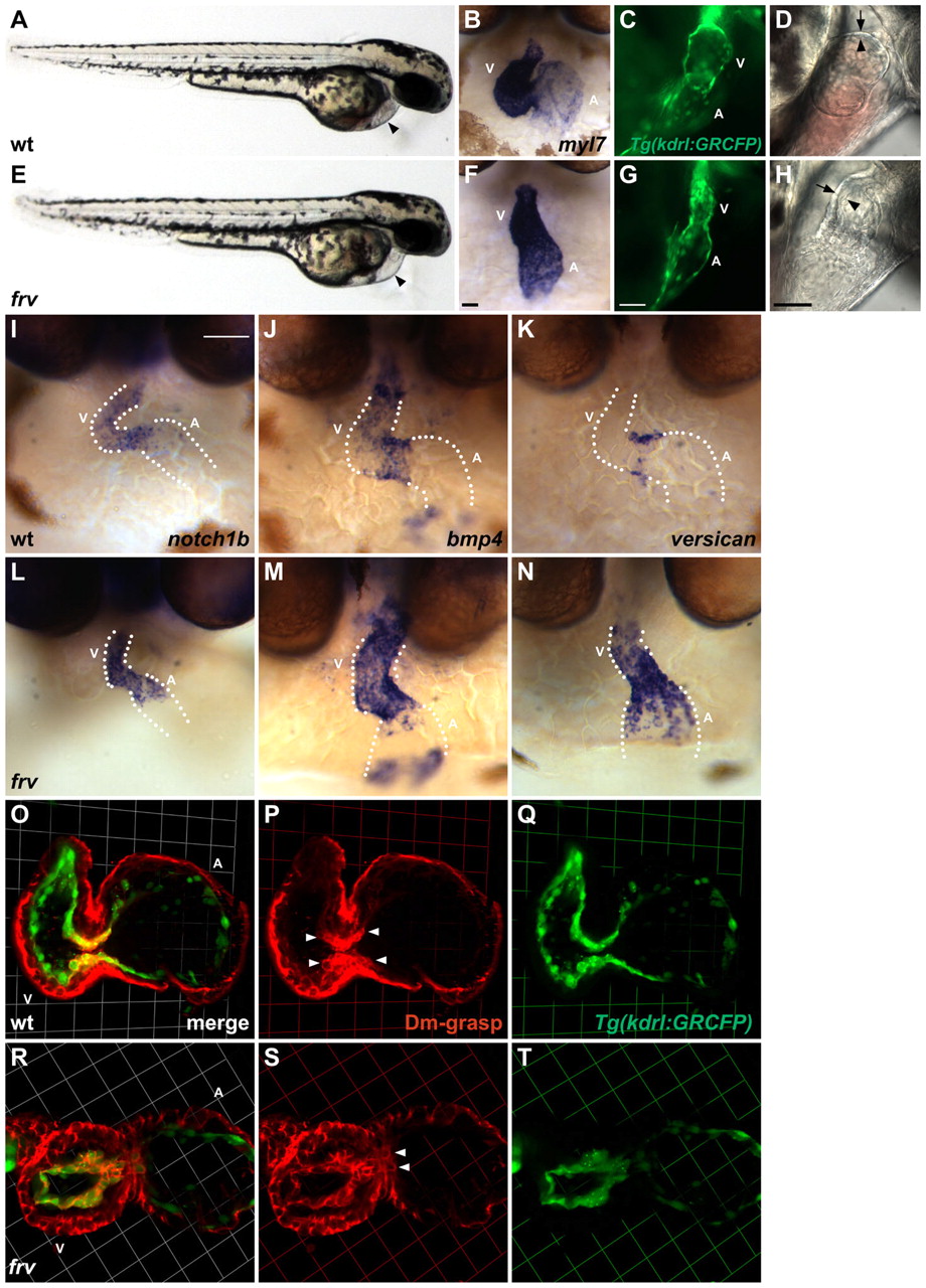
The frv mutation disrupts myocardial and endocardial morphogenesis. (A,E) Lateral views of wild-type (wt) and frv mutant zebrafish embryos at 48 hpf reveal mild pericardial edema in frv mutants (arrowheads). Other than their cardiac defects, frv mutants appear morphologically normal. (B,F) Frontal views depict myl7 expression in wild type (B) and in frv mutants (F) at 48 hpf. (C,G) Lateral views showing endocardial expression of Tg(kdrl:GRCFP) in wild type (C) and in frv mutants (G) at 52 hpf. (D,H) Lateral views at 52 hpf depict the close juxtaposition of the ventricular endocardium (arrowhead) and myocardium (arrow) in wild type (D) and their greater separation in frv mutants (H). (I-N) Frontal views depict expression of atrioventricular canal (AVC) markers in wild type (I-K) and in frv mutants (L-N) at 48 hpf. Dotted lines outline the chambers flanking the AVC. (O-T) Three-dimensional projections of selected confocal sections of wild-type (O-Q) and frv mutant (R-T) hearts expressing Tg(kdrl:GRCFP) (green) in the endocardium at 57 hpf. Immunofluorescence reveals Dm-grasp (red) throughout the myocardium and in the AVC endocardium (arrowheads). In frv mutants, Dm-grasp is also seen ectopically in the ventricular endocardium. Grids are 23 μm per segment. A, atrium; V, ventricle. Scale bars: 50 μm.