Fig. 4
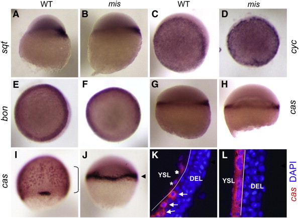
Defects in endoderm-specific gene expression in maternally mutant mis embryos occur in downstream steps within the pathway. Wild-type (A,C,E,G,I,K) and mis mutant (B,D,F,H,J,L) embryos assayed for expressed transcripts encoding endoderm signaling components, including the nodal signals squint (A,B) and cyclops (C,D), and the transcription factors bon/mixer (E,F) and casanova/sox32 (cas, G-L). Expression patterns for squint, cyclops, and bon appear similar in wild-type and mis mutant embryos. cas becomes induced in the YSL at 5 h.p.f. in both wild-type and mis mutants (G,H). By 8 h.p.f., endodermal cells extending into the hypoblast layer express cas in a “salt and pepper” distribution in wild-type (I, bracket). However, cas expression in mis mutant embryos at this stage is restricted to the blastoderm margin (J, arrowhead). (K,L) Closer analysis of cas gene expression in the YSL and DEL was carried out by FISH with a cas antisense probe (red) and DAPI labeling (blue), followed by confocal microscopy. The YSL layer can be distinguished from the DEL by the less dense arrangement of its nuclei and their larger size, and a white line has been drawn between the two layers. In wild-type embryos at this stage (K), expression of cas in the YSL is subsiding (asterisks indicate YSL nuclei associated with cas mRNA), while strong expression has begun in interspersed DEL cells tightly adjoining the YSL (arrows), the presumed endodermal precursors. On the other hand, mis embryos (L) show abnormally strong and persistent cas expression in the YSL and very few, if any, cas-expressing cells in the DEL. Stages are 5 h.p.f. (A–D, G,H), 6 h.p.f. (E,F), and 8 h.p.f. (I–L). (A,B,G,H) are lateral views with dorsal to the right, when identifiable. (C–F) are animal pole views (expression in embryos corresponding to these panels is largely symmetric and slight apparent asymmetries in the embryos shown are caused by photographing artifacts). (I,J) are dorsal views. (K,L) are optical sections through the embryonic blastoderm at about 1/3 of the way between the margin and the animal pole.
Reprinted from Developmental Biology, 353(2), Putiri, E., and Pelegri, F., The zebrafish maternal-effect gene mission impossible encodes the DEAH-box helicase Dhx16 and is essential for the expression of downstream endodermal genes, 275-289, Copyright (2011) with permission from Elsevier. Full text @ Dev. Biol.

