Fig. 2
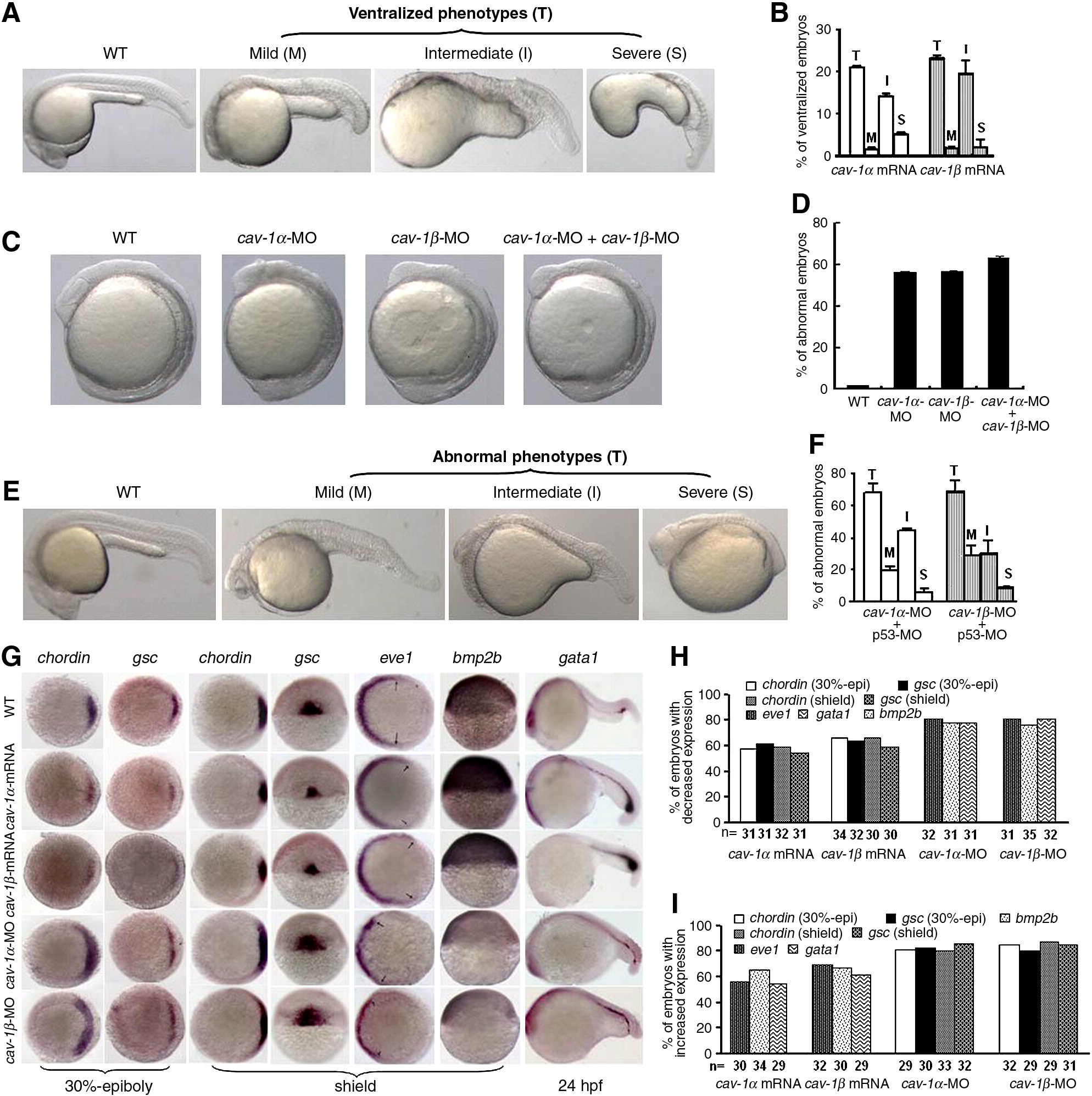
Fig. 2 Effects of cav-1 overexpression and knockdown on zebrafish dorsoventral patterning. (A) Overexpression of cav-1 ventralizes zebrafish embryos. Embryos at one-cell stage were injected with 300 pg cav-1α or -1β mRNA. Wild-type (WT) embryos was used as the control. Total ventralized embryos (T) were divided into three groups: mild (M), intermediate (I) and severe (S). (B) Percentage of ventralized embryos for each group in (A). Data represent mean ± S.D. from three independent experiments. (C) Knockdown of cav-1 led to weakly dorsalized embryos with early tailbud protrusion at 12 hpf. Embryos at one-cell stage were injected with 10 ng cav-1α-MO, 10 ng cav-1β-MO, or 5 ng cav-1α-MO plus 5 ng cav-1β-MO. (D) Percentage of abnormal embryos at 12 hpf for each group in (C). (E) Knockdown of cav-1 led to abnormal embryos at 24 hpf. Embryos at one-cell stage were injected with 1 ng p53 MO plus 10 ng cav-1α-MO or -1β-MO. Total number of abnormal embryos (T) at 24 hpf was divided into three groups: mild (M), intermediate (I) and severe (S). (F) Percentage of abnormal embryos at 24 hpf for each group in (E). Data represent mean ± S.D. from three independent experiments. (G) Effects of cav-1 overexpression and knockdown on expression patterns of dorsoventral marker genes (indicated on the top). Developmental stages are shown at the bottom. Lateral views of embryos at shield stage with the dorsal toward the right for bmp2b. Animal pole views of embryos at shield stage with the dorsal toward the right for chordin, eve1 and gsc at 30% epiboly stage. Dorsal views of embryos at shield stage with the animal pole toward the top for gsc at shield stage. Lateral views of embryos at 24 hpf with the anterior to the left for gata1. Arrows point to the margins of expressed eve1. (H–I) Total number of embryos examined (indicated at the bottom) and percentage of embryos with decreased or increased expression of dorsoventral marker genes in (G).
Reprinted from Developmental Biology, 344(1), Mo, S., Wang, L., Li, Q., Li, J., Li, Y., Thannickal, V.J., and Cui. Z., Caveolin-1 regulates dorsoventral patterning through direct interaction with beta-catenin in zebrafish, 210-223, Copyright (2010) with permission from Elsevier. Full text @ Dev. Biol.

