Fig. 2
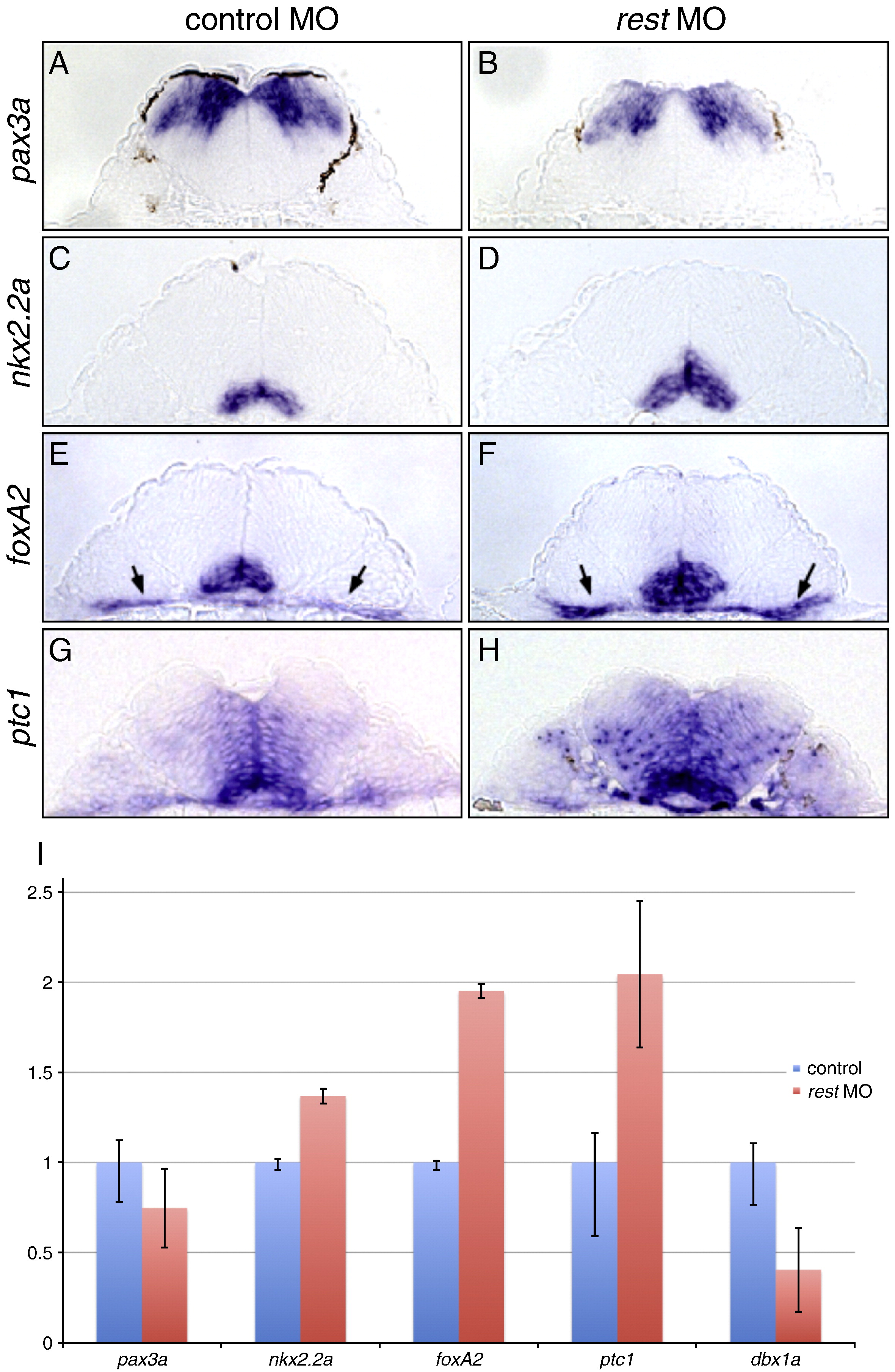
Fig. 2 The neural tube is ventralized in Rest knockdown embryos. Transverse sections of 29hpf (A, B) or 26hpf (C–H) wild-type embryos processed for RNA in situ hybridization and sectioned at the level of the hindbrain at anterior rhombomere 4. Control (A, C, E, G) and rest MO injected (B, D, F, H) embryos stained with antisense probes for Hh response genes pax3a, nkx2.2a, foxA2 and ptc1. pax3a expression (A, B) is reduced in rest morphants, while expression of nkx2.2a (C, D), foxA2 (E, F) and ptc1 (G, H) is expanded compared to stage matched control embryos. This suggests that Rest represses Hh signaling. Arrows (E, F) mark pharyngeal endoderm. (I) qPCR analysis on 29hpf control (red bars) and stage matched rest morphant (blue bars) cDNA for markers shown in A–H, plus class I gene dbx1a. Overall levels of class II Hh target genes nkx2.2a, foxA2, and ptc1 are increased while class I genes pax3a and dbx1a levels are reduced.
Reprinted from Developmental Biology, 340(2), Gates, K.P., Mentzer, L., Karlstrom, R.O., and Sirotkin, H.I., The transcriptional repressor REST/NRSF modulates hedgehog signaling, 293-305, Copyright (2010) with permission from Elsevier. Full text @ Dev. Biol.

