
Fig. 2
- ID
- ZDB-IMAGE-091016-25
- Genes
- Antibodies
- Publication
- Anderson et al., 2009 - Loss of Dnmt1 catalytic activity reveals multiple roles for DNA methylation during pancreas development and regeneration
- All Figures
- Figures for Anderson et al., 2009
|
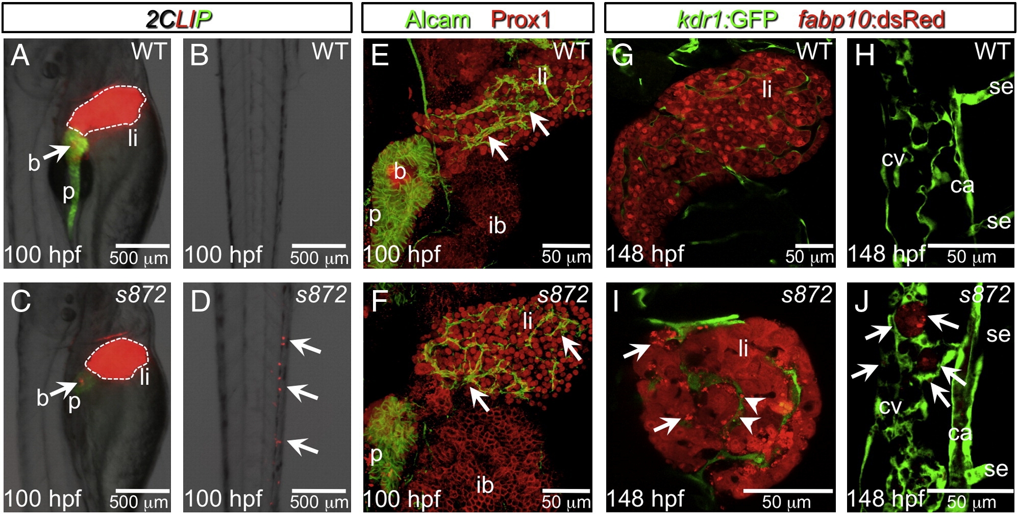
Fig. 2 Liver degeneration in dandelion (ddn) mutants. (A–D) Lateral aspect of trunk (A, C) and tail (B, D) regions of 100 hpf WT (A, B) and ddn mutant (C, D) larvae in the 2CLIP transgenic background. The liver is smaller in ddn mutants relative to WT, and cell fragments emitting dsRed fluorescence, derived from the liver, aggregate in clusters in the tail (D, arrows). (E, F) 3-D projections of confocal stacks showing Alcam and Prox1 antibody staining of 100 hpf WT (E) and ddn mutants (F). Initial specification, morphogenesis, and differentiation of the liver appear to be unaffected. Intrahepatic ductal network is indicated by arrows. (G–J) Confocal slices through the liver (G, I) and tail vasculature (H, J) of Tg(kdrl:GFP)s843; Tg(fabp10:dsRed)gz12 WT (G, H) and ddn mutant (I, J) larvae at 6 dpf. Although never present in WT, bright dsRed+ degenerating hepatocyte fragments are observed throughout the ddn mutant liver (I, arrows) and within the hepatic vasculature (I, arrowheads). These particles accumulate in the network of the caudal vein (cv; J, arrows). Other abbreviations: ca, caudal aorta; se, intersegmental vessel; ib, intestinal bulb; p, pancreas; li, liver; b, beta cells.
Reprinted from Developmental Biology, 334(1), Anderson, R.M., Bosch, J.A., Goll, M.G., Hesselson, D., Dong, P.D., Shin, D., Chi, N.C., Shin, C.H., Schlegel, A., Halpern, M., and Stainier, D.Y., Loss of Dnmt1 catalytic activity reveals multiple roles for DNA methylation during pancreas development and regeneration, 213-223, Copyright (2009) with permission from Elsevier. Full text @ Dev. Biol.