
Fig. S4
- ID
- ZDB-IMAGE-090804-95
- Genes
- Antibodies
- Publication
- Nikolaou et al., 2009 - Lunatic fringe promotes the lateral inhibition of neurogenesis
- All Figures
- Figures for Nikolaou et al., 2009
|
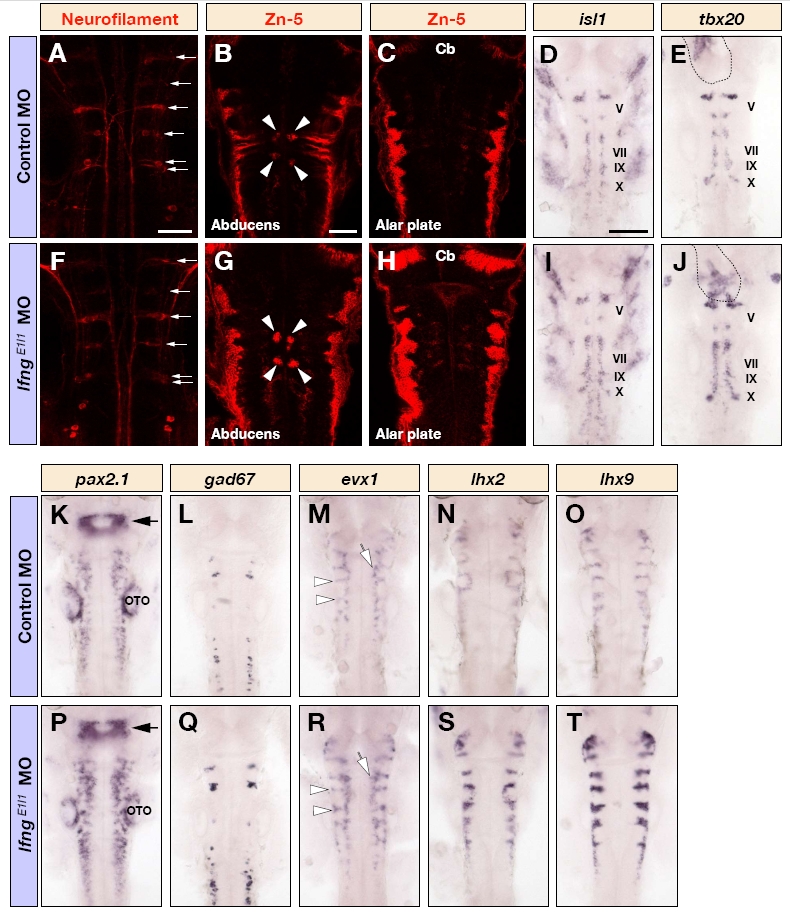
Fig. S4 Many neuronal subtypes are affected following knockdown of lfng. (A-T) Dorsal views of the hindbrain of control MO (A-E,K-O) and lfngE1I1 MO (F-J,P-T) injected embryos at 26 hpf (D,E,I,J,L,Q), 30 hpf (K,M,N,O,P,R,S,T) and 48 hpf (A-C,F-H) following immunostaining for neurofilament (A,F) or Zn-5 (B,C,G,H), or detection of isl1 (D,I), tbx20 (E,J), pax2.1 (K,P), gad67 (L,Q), evx1 (M,R), lxh2 (N,S) or lhx9 (O,T) mRNA. There is an increase in the number of somatic motor neurons (arrowheads in B and G), dorsal hindbrain neurons (G,H), branchiomotor neurons (D,E,I,J), various interneuronal populations (K,L,M,P,Q,R) and commissural neurons (M,N,O,R,S,T). Note that reticulospinal neurons are not affected (arrows in A and F). The outline in E and J indicates the expression of tbx20 in the developing heart. The arrows in K and P indicate the mid-hindbrain boundary. The open arrows and arrowheads in M and R show the expression of evx1 in putative interneurons and commissural neurons, respectively. Cb, cerebellum; OTO, otocyst. Scale bars: 50 μm in A and B for A-C,F-H; in (B); 100 μm in D for D,E, I-T.