
Fig. 3
- ID
- ZDB-IMAGE-080829-44
- Genes
- Antibodies
- Publication
- Hsu et al., 2006 - Mosaic Eyes is a novel component of the Crumbs complex and negatively regulates photoreceptor apical size
- All Figures
- Figures for Hsu et al., 2006
|
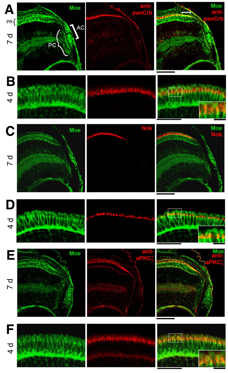
Fig. 3 Localization of Moe, panCrb, Nok and aPKCλ in the retina. (A,B) Colocalization of guinea pig anti-Moe and panCrb labeling in the peripheral region in 7 dpf eye (A) and photoreceptor layer at 4 dpf (B). AC, anterior chamber; PC, proliferating cells; PR, photoreceptors. (C,D) Colocalization of guinea pig anti-Moe and anti-Nok labeling in the peripheral region in 7 dpf eye (C) and photoreceptor layer at 4 dpf (D). (E,F) Colocalization of guinea pig anti-Moe and anti-aPKCλ labeling in the peripheral region in 7 dpf eye (E) and photoreceptor layer at 4 dpf (F). Insets are magnification of boxed areas. Scale bars: 20 μm; 10 μm in insets.