
Fig. 3
- ID
- ZDB-IMAGE-080828-8
- Genes
- Publication
- Dee et al., 2008 - Sox3 regulates both neural fate and differentiation in the zebrafish ectoderm
- All Figures
- Figures for Dee et al., 2008
|
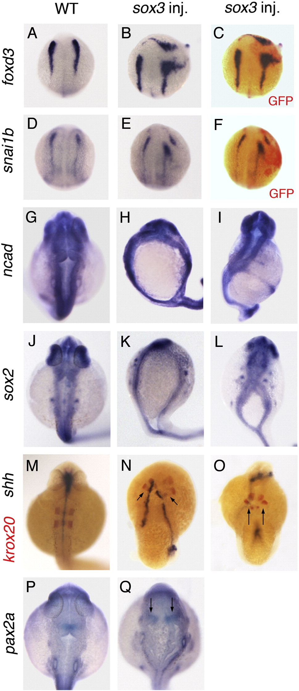
Fig. 3 Over-expression of Sox3 can cause duplications of the CNS. (A–F) Dorsal view of embryos at the 3 somite stage. (A, D) Control wild-type embryos showing expression of neural crest markers foxd3 (A) and snai1b (D) flanking the neural plate. (B, E) Embryos injected with sox3 mRNA (and GFP mRNA lineage tracer) at the 16–32 cell stage can generate a partial second axis flanked by foxd3 (B, 4/30 embryos) or snai1b (E, 2/39) expression. Immunostaining for GFP (red colour) shows this to correspond to the injected region (C, F). (G–Q) Embryos viewed dorsally aged 24 hpf. (G, J, M, P) Wild-type control embryos. (G–L) In situ hybridisation for the expression of neural markers ncad (G–I) and sox2 (J–L) reveals that injection of sox3 mRNA at 16–32 cell stage can cause duplications of the CNS in the head (H, 2/21, K, 5/47) or tail (I, 3/21, L, 14/47). (M–Q) Duplications are also seen of the hindbrain marker krox20 (M–O, red colour, arrows indicate duplication, 8/43), the floor plate marker shha (M–O, blue colour) and pax2a which is expressed at the midbrain/hindbrain boundary (arrows, P, Q).
Reprinted from Developmental Biology, 320(1), Dee, C.T., Hirst, C.S., Shih, Y.H., Tripathi, V.B., Patient, R.K., and Scotting, P.J., Sox3 regulates both neural fate and differentiation in the zebrafish ectoderm, 289-301, Copyright (2008) with permission from Elsevier. Full text @ Dev. Biol.